 Movie
Movie Controller


+ Open data

- Basic information
| Entry | Database: PDB / ID: 6ygi | ||||||||||||
|---|---|---|---|---|---|---|---|---|---|---|---|---|---|
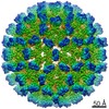
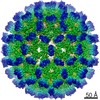
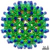
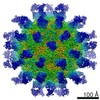
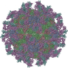
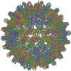
| Title | Duck hepatitis B virus capsid Mutant R124E_delta78-122 | ||||||||||||
 Components Components | Capsid protein,Capsid protein | ||||||||||||
 Keywords Keywords | VIRUS LIKE PARTICLE / duck hepatitis B core protein / extension domain / spike / slowly folding | ||||||||||||
| Function / homology |  Function and homology information Function and homology informationmicrotubule-dependent intracellular transport of viral material towards nucleus / T=4 icosahedral viral capsid / viral penetration into host nucleus / host cell cytoplasm / symbiont entry into host cell / structural molecule activity / DNA binding / RNA bindingSimilarity search - Function | ||||||||||||
| Biological species |  Hepatitis B virus duck/DHBV-16 Hepatitis B virus duck/DHBV-16 | ||||||||||||
| Method | ELECTRON MICROSCOPY / single particle reconstruction / cryo EM / Resolution: 3 Å | ||||||||||||
 Authors Authors | Makbul, C. / Bottcher, B. | ||||||||||||
| Funding support |  Germany, 3items Germany, 3items
| ||||||||||||
 Citation Citation | Journal: Elife / Year: 2020 Title: Slowly folding surface extension in the prototypic avian hepatitis B virus capsid governs stability. Authors: Cihan Makbul / Michael Nassal / Bettina Böttcher / Abstract: Hepatitis B virus (HBV) is an important but difficult to study human pathogen. Most basics of the hepadnaviral life-cycle were unraveled using duck HBV (DHBV) as a model although DHBV has a capsid ...Hepatitis B virus (HBV) is an important but difficult to study human pathogen. Most basics of the hepadnaviral life-cycle were unraveled using duck HBV (DHBV) as a model although DHBV has a capsid protein (CP) comprising ~260 rather than ~180 amino acids. Here we present high-resolution structures of several DHBV capsid-like particles (CLPs) determined by electron cryo-microscopy. As for HBV, DHBV CLPs consist of a dimeric α-helical frame-work with protruding spikes at the dimer interface. A fundamental new feature is a ~ 45 amino acid proline-rich extension in each monomer replacing the tip of the spikes in HBV CP. In vitro, folding of the extension takes months, implying a catalyzed process in vivo. DHBc variants lacking a folding-proficient extension produced regular CLPs in bacteria but failed to form stable nucleocapsids in hepatoma cells. We propose that the extension domain acts as a conformational switch with differential response options during viral infection. | ||||||||||||
| History |
|
- Structure visualization
| Movie |
Movie viewer |
|---|---|
| Structure viewer | Molecule: MolmilJmol/JSmol |
- Downloads & links
-Download
| PDBx/mmCIF format | 6ygi.cif.gz | 222 KB | Display | PDBx/mmCIF format |
|---|---|---|---|---|
| PDB format | pdb6ygi.ent.gz | 184.5 KB | Display | PDB format |
| PDBx/mmJSON format | 6ygi.json.gz | Tree view | PDBx/mmJSON format | |
| Others |  Other downloads Other downloads |
-Validation report
| Arichive directory | https://data.pdbj.org/pub/pdb/validation_reports/yg/6ygiftp://data.pdbj.org/pub/pdb/validation_reports/yg/6ygi | HTTPS FTP |
|---|
-Related structure data
| Related structure data | 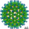 10803MC  6yghC C: citing same article ( M: map data used to model this data |
|---|---|
| Similar structure data |


-Links
 PDBj
PDBj

- Assembly
| Deposited unit | 
|
|---|---|
| 1 | x 60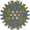
|
-Components
| #1: Protein | / Core antigen / Core protein / HBcAg Mass: 25495.988 Da / Num. of mol.: 6 Source method: isolated from a genetically manipulated source Details: deletion mutant with extension domain (78-122) replaced by flexible linker GGSGG additional point mutation R124E,deletion mutant with extension domain (78-122) replaced by flexible linker ...Details: deletion mutant with extension domain (78-122) replaced by flexible linker GGSGG additional point mutation R124E,deletion mutant with extension domain (78-122) replaced by flexible linker GGSGG additional point mutation R124E Source: (gene. exp.) Hepatitis B virus duck/DHBV-16 / Production host:  Escherichia coli BL21(DE3) (bacteria) / References: UniProt: P0C6J7 Escherichia coli BL21(DE3) (bacteria) / References: UniProt: P0C6J7 |
|---|
-Experimental details
-Experiment
| Experiment | Method: ELECTRON MICROSCOPY |
|---|---|
| EM experiment | Aggregation state: PARTICLE / 3D reconstruction method: single particle reconstruction |
- Sample preparation
| Component | Name: Hepatitis B virus duck/DHBV-16 / Type: VIRUS / Entity ID: all / Source: RECOMBINANT | ||||||||||||||||||||||||||||||
|---|---|---|---|---|---|---|---|---|---|---|---|---|---|---|---|---|---|---|---|---|---|---|---|---|---|---|---|---|---|---|---|
| Molecular weight | Value: 7 MDa / Experimental value: NO | ||||||||||||||||||||||||||||||
| Source (natural) | Organism: Hepatitis B virus duck/DHBV-16 | ||||||||||||||||||||||||||||||
| Source (recombinant) | Organism: Escherichia coli BL21(DE3) (bacteria) | ||||||||||||||||||||||||||||||
| Details of virus | Empty: NO / Enveloped: NO / Isolate: SPECIES / Type: VIRUS-LIKE PARTICLE | ||||||||||||||||||||||||||||||
| Natural host | Organism: Anas platyrhynchos | ||||||||||||||||||||||||||||||
| Virus shell | Name: duck Hepatitis B virus capsid / Diameter: 370 nm / Triangulation number (T number): 4 | ||||||||||||||||||||||||||||||
| Buffer solution | pH: 7.5 | ||||||||||||||||||||||||||||||
| Buffer component |
| ||||||||||||||||||||||||||||||
| Specimen | Conc.: 3 mg/ml / Embedding applied: NO / Shadowing applied: NO / Staining applied: NO / Vitrification applied: YES | ||||||||||||||||||||||||||||||
| Specimen support | Grid material: COPPER / Grid mesh size: 400 divisions/in. / Grid type: Quantifoil R1.2/1.3 | ||||||||||||||||||||||||||||||
| Vitrification | Instrument: FEI VITROBOT MARK IV / Cryogen name: ETHANE / Humidity: 100 % / Chamber temperature: 277 K Details: For the vitrification, grids (400 mesh copper grids (type R 1.2/1.3. Quantifoil Micro Tools, Jena/Germany) were rendered hydrophilic by glow discharging in air at a pressure of 29 Pa for 2 ...Details: For the vitrification, grids (400 mesh copper grids (type R 1.2/1.3. Quantifoil Micro Tools, Jena/Germany) were rendered hydrophilic by glow discharging in air at a pressure of 29 Pa for 2 minutes at medium power with a Plasma Cleaner (model PDC-002. Harrick Plasma Ithaca, NY/USA). Then, 3.5 ul of DHBc solution was pipetted onto the grids and they were plunge frozen in liquid ethane with a Vitrobot mark IV (FEI-Thermo Fisher Scientific). The settings for the Vitrobot were 3s blot time, 45 s wait time, blot force 0 at a temperature of 4 C and 100 % humidity |
- Electron microscopy imaging
| Experimental equipment |  Model: Titan Krios / Image courtesy: FEI Company |
|---|---|
| Microscopy | Model: FEI TITAN KRIOS |
| Electron gun | Electron source: FIELD EMISSION GUN / Accelerating voltage: 300 kV / Illumination mode: FLOOD BEAM |
| Electron lens | Mode: BRIGHT FIELDBright-field microscopy / Nominal magnification: 75000 X / Calibrated defocus min: 500 nm / Calibrated defocus max: 1300 nm / Cs: 2.7 mm / C2 aperture diameter: 70 µm / Alignment procedure: COMA FREE |
| Specimen holder | Cryogen: NITROGEN / Specimen holder model: FEI TITAN KRIOS AUTOGRID HOLDER |
| Image recording | Average exposure time: 54 sec. / Electron dose: 60 e/Å2 / Detector mode: COUNTING / Film or detector model: FEI FALCON III (4k x 4k) / Num. of grids imaged: 1 / Num. of real images: 1496 Details: movie mode, 2 images per hole, 40 fractions per movie |
| Image scans | Width: 4096 / Height: 4096 |
- Processing
| Software | Name: PHENIX / Version: 1.16_3549: / Classification: refinement | |||||||||||||||||||||||||||||||||||||||||||||||||||||||
|---|---|---|---|---|---|---|---|---|---|---|---|---|---|---|---|---|---|---|---|---|---|---|---|---|---|---|---|---|---|---|---|---|---|---|---|---|---|---|---|---|---|---|---|---|---|---|---|---|---|---|---|---|---|---|---|---|
| EM software |
| |||||||||||||||||||||||||||||||||||||||||||||||||||||||
| Image processing | Details: Micrographs were dose weighted and motion corrected with Motioncor2. The ctf was determined with ctffind4 | |||||||||||||||||||||||||||||||||||||||||||||||||||||||
| CTF correction | Type: PHASE FLIPPING AND AMPLITUDE CORRECTION | |||||||||||||||||||||||||||||||||||||||||||||||||||||||
| Particle selection | Num. of particles selected: 74760 Details: particles were automatically selected using 2 D class averages as template | |||||||||||||||||||||||||||||||||||||||||||||||||||||||
| Symmetry | Point symmetry: I (icosahedral) | |||||||||||||||||||||||||||||||||||||||||||||||||||||||
| 3D reconstruction | Resolution: 3 Å / Resolution method: FSC 0.143 CUT-OFF / Num. of particles: 44498 / Algorithm: FOURIER SPACE / Num. of class averages: 1 / Symmetry type: POINT | |||||||||||||||||||||||||||||||||||||||||||||||||||||||
| Atomic model building | Protocol: RIGID BODY FIT / Space: REAL | |||||||||||||||||||||||||||||||||||||||||||||||||||||||
| Refine LS restraints |
|
