 Movie
Movie Controller


+ Open data

- Basic information
| Entry | Database: PDB / ID: 6ye7 | ||||||
|---|---|---|---|---|---|---|---|
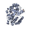
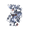
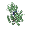
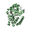
| Title | E.coli's Putrescine receptor PotF complexed with Cadaverine | ||||||
 Components Components | Putrescine-binding periplasmic protein | ||||||
 Keywords Keywords |  TRANSPORT PROTEIN / Periplasmic binding protein / E.coli / Cadaverine TRANSPORT PROTEIN / Periplasmic binding protein / E.coli / Cadaverine | ||||||
| Function / homology |  Function and homology information Function and homology informationputrescine transport / putrescine binding / ATP-binding cassette (ABC) transporter complex, substrate-binding subunit-containing / outer membrane-bounded periplasmic space / membraneSimilarity search - Function | ||||||
| Biological species |  Escherichia coli (E. coli) Escherichia coli (E. coli) | ||||||
| Method | X-RAY DIFFRACTION / SYNCHROTRON / MOLECULAR REPLACEMENT / Resolution: 1.6 Å | ||||||
 Authors Authors | Shanmugaratnam, S. / Kroeger, P. / Hocker, B. | ||||||
| Funding support |  Germany, 1items Germany, 1items
| ||||||
 Citation Citation | Journal: Structure / Year: 2021 Title: A comprehensive binding study illustrates ligand recognition in the periplasmic binding protein PotF. Authors: Kroger, P. / Shanmugaratnam, S. / Ferruz, N. / Schweimer, K. / Hocker, B. | ||||||
| History |
|
- Structure visualization
| Structure viewer | Molecule: MolmilJmol/JSmol |
|---|
- Downloads & links
-Download
| PDBx/mmCIF format | 6ye7.cif.gz | 512.9 KB | Display | PDBx/mmCIF format |
|---|---|---|---|---|
| PDB format | pdb6ye7.ent.gz | 351.7 KB | Display | PDB format |
| PDBx/mmJSON format | 6ye7.json.gz | Tree view | PDBx/mmJSON format | |
| Others |  Other downloads Other downloads |
-Validation report
| Arichive directory | https://data.pdbj.org/pub/pdb/validation_reports/ye/6ye7ftp://data.pdbj.org/pub/pdb/validation_reports/ye/6ye7 | HTTPS FTP |
|---|
-Related structure data
| Related structure data |  6ye0C  6ye6C  6ye8C  6yebC  6yecC  6yedC  1a99S C: citing same article ( S: Starting model for refinement |
|---|---|
| Similar structure data |


-Links
 PDBj
PDBj


- Assembly
| Deposited unit | 
| ||||||||||||||||||||||||||||||||||||||||||||||||||||||||||||||||||||||
|---|---|---|---|---|---|---|---|---|---|---|---|---|---|---|---|---|---|---|---|---|---|---|---|---|---|---|---|---|---|---|---|---|---|---|---|---|---|---|---|---|---|---|---|---|---|---|---|---|---|---|---|---|---|---|---|---|---|---|---|---|---|---|---|---|---|---|---|---|---|---|---|
| 1 | 
| ||||||||||||||||||||||||||||||||||||||||||||||||||||||||||||||||||||||
| 2 | 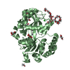
| ||||||||||||||||||||||||||||||||||||||||||||||||||||||||||||||||||||||
| Unit cell |
| ||||||||||||||||||||||||||||||||||||||||||||||||||||||||||||||||||||||
| Noncrystallographic symmetry (NCS) | NCS domain:
NCS domain segments:
|