 Movie
Movie Controller

[English] 日本語
 Yorodumi
- PDB-6y8j: Crystal structure of the apo form of a quaternary ammonium Rieske... -
+ Open data

- Basic information
| Entry | Database: PDB / ID: 6y8j | ||||||
|---|---|---|---|---|---|---|---|
| Title | Crystal structure of the apo form of a quaternary ammonium Rieske monooxygenase CntA | ||||||
 Components Components | Carnitine monooxygenase oxygenase subunit | ||||||
 Keywords Keywords |  OXIDOREDUCTASE / Apo / Rieske / Iron-Sulphur Cluster OXIDOREDUCTASE / Apo / Rieske / Iron-Sulphur Cluster | ||||||
| Function / homology |  Function and homology information Function and homology informationcarnitine monooxygenase / carnitine metabolic process / oxidoreductase activity, acting on paired donors, with incorporation or reduction of molecular oxygen, NAD(P)H as one donor, and incorporation of one atom of oxygen / dioxygenase activity / 2 iron, 2 sulfur cluster binding / iron ion bindingSimilarity search - Function | ||||||
| Biological species |  Acinetobacter baumannii (bacteria) Acinetobacter baumannii (bacteria) | ||||||
| Method | X-RAY DIFFRACTION / SYNCHROTRON / MOLECULAR REPLACEMENT / Resolution: 2.05 Å | ||||||
 Authors Authors | Quareshy, M. / Shanmugam, M. / Bugg, T.D. / Cameron, A. / Chen, Y. | ||||||
| Funding support |  United Kingdom, 1items United Kingdom, 1items
| ||||||
 Citation Citation | Journal: J.Biol.Chem. / Year: 2020 Title: Structural basis of carnitine monooxygenase CntA substrate specificity, inhibition, and intersubunit electron transfer. Authors: Quareshy, M. / Shanmugam, M. / Townsend, E. / Jameson, E. / Bugg, T.D.H. / Cameron, A.D. / Chen, Y. | ||||||
| History |
|
- Structure visualization
| Structure viewer | Molecule: MolmilJmol/JSmol |
|---|
- Downloads & links
-Download
| PDBx/mmCIF format | 6y8j.cif.gz | 160.5 KB | Display | PDBx/mmCIF format |
|---|---|---|---|---|
| PDB format | pdb6y8j.ent.gz | 124.3 KB | Display | PDB format |
| PDBx/mmJSON format | 6y8j.json.gz | Tree view | PDBx/mmJSON format | |
| Others |  Other downloads Other downloads |
-Validation report
| Arichive directory | https://data.pdbj.org/pub/pdb/validation_reports/y8/6y8jftp://data.pdbj.org/pub/pdb/validation_reports/y8/6y8j | HTTPS FTP |
|---|
-Related structure data
| Related structure data |  6y8sC 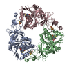 6y9dC  6zgpC 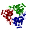 3vcpS S: Starting model for refinement C: citing same article ( |
|---|---|
| Similar structure data |


-Links
 PDBj
PDBj


- Assembly
| Deposited unit | 
| ||||||||
|---|---|---|---|---|---|---|---|---|---|
| 1 | 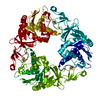
| ||||||||
| Unit cell |
|
-Components
| #1: Protein | Mass: 44780.172 Da / Num. of mol.: 1 Source method: isolated from a genetically manipulated source Source: (gene. exp.) Acinetobacter baumannii (bacteria)Gene: antA_2, antA_1, antA_3, A7M79_02670, A7M90_13970, ABUW_3074, B4R90_07590, B9X95_06095, BGC29_09330, C2U32_18540, C3415_14505, CBI29_00874, CHQ89_11265, CPI82_11190, CSB70_0522, DLI75_01970, ...Gene: antA_2, antA_1, antA_3, A7M79_02670, A7M90_13970, ABUW_3074, B4R90_07590, B9X95_06095, BGC29_09330, C2U32_18540, C3415_14505, CBI29_00874, CHQ89_11265, CPI82_11190, CSB70_0522, DLI75_01970, DOL94_04925, DVA79_16365, E2533_13315, E2536_16135, E5294_15630, E5979_13670, EA685_07170, EA686_01565, EA706_03020, EA722_03860, EA746_003300, EWO92_12480, EWO96_16565, EWP49_15025, FD887_09300, FD913_14110, FJU36_15000, FJU42_16200, FJU76_14830, FJU79_08840, FJU87_10695, FJV14_20515, LV38_02893, NCTC13305_01609, SAMEA104305283_02985, SAMEA104305351_01970 Plasmid: pET-28 a (+) / Production host: Escherichia coli BL21(DE3) (bacteria) / Variant (production host): pLysS / References: UniProt: A0A059ZPP5, carnitine monooxygenase |
|---|---|
| #2: Chemical | ChemComp-FES / Iron–sulfur cluster  Mass: 175.820 Da / Num. of mol.: 1 / Source method: obtained synthetically / Formula: Fe2S2 / Feature type: SUBJECT OF INVESTIGATION Mass: 175.820 Da / Num. of mol.: 1 / Source method: obtained synthetically / Formula: Fe2S2 / Feature type: SUBJECT OF INVESTIGATION |
| #3: Water | ChemComp-HOH / Water Mass: 18.015 Da / Num. of mol.: 66 / Source method: isolated from a natural source / Formula: H2O Mass: 18.015 Da / Num. of mol.: 66 / Source method: isolated from a natural source / Formula: H2O |
| Has ligand of interest | Y |
-Experimental details
-Experiment
| Experiment | Method: X-RAY DIFFRACTION / Number of used crystals: 1 |
|---|
- Sample preparation
| Crystal | Density Matthews: 2.18 Å3/Da / Density % sol: 43.64 % / Description: Red Hexagonal Crystals |
|---|---|
| Crystal grow | Temperature: 295 K / Method: vapor diffusion, hanging drop / pH: 7.6 / Details: 10mM HEPES, 20% PEG3350, 0.2M NaSCN, 0.5mM TCEP / PH range: 7.0 - 7.6 |
-Data collection
| Diffraction | Mean temperature: 100 K / Serial crystal experiment: N | ||||||||||||||||||||||||||||||
|---|---|---|---|---|---|---|---|---|---|---|---|---|---|---|---|---|---|---|---|---|---|---|---|---|---|---|---|---|---|---|---|
| Diffraction source | Source: SYNCHROTRON / Site: Diamond / Beamline: I24 / Wavelength: 1.7397 Å | ||||||||||||||||||||||||||||||
| Detector | Type: DECTRIS PILATUS3 6M / Detector: PIXEL / Date: Feb 10, 2019 | ||||||||||||||||||||||||||||||
| Radiation | Protocol: SINGLE WAVELENGTH / Monochromatic (M) / Laue (L): M / Scattering type: x-ray | ||||||||||||||||||||||||||||||
| Radiation wavelength | Wavelength: 1.7397 Å / Relative weight: 1 | ||||||||||||||||||||||||||||||
| Reflection | Resolution: 2.05→45.8 Å / Num. obs: 24733 / % possible obs: 99.3 % / Redundancy: 9.1 % / CC1/2: 0.994 / Rmerge(I) obs: 0.195 / Rpim(I) all: 0.066 / Rrim(I) all: 0.206 / Net I/σ(I): 7.4 / Num. measured all: 225029 / Scaling rejects: 544 | ||||||||||||||||||||||||||||||
| Reflection shell | Diffraction-ID: 1
|
- Processing
| Software |
| ||||||||||||||||||||||||||||||||||||||||||||||||||||||||||||||||||||||||||||||||||||||||||||||||||||||||||||||||||||||||||||||||||||||||||||||||||||||||||||||||||||||||||||||||||||||||||||||||||||||||||||||||||||||||||||||||||||||||||||||||||||||||||
|---|---|---|---|---|---|---|---|---|---|---|---|---|---|---|---|---|---|---|---|---|---|---|---|---|---|---|---|---|---|---|---|---|---|---|---|---|---|---|---|---|---|---|---|---|---|---|---|---|---|---|---|---|---|---|---|---|---|---|---|---|---|---|---|---|---|---|---|---|---|---|---|---|---|---|---|---|---|---|---|---|---|---|---|---|---|---|---|---|---|---|---|---|---|---|---|---|---|---|---|---|---|---|---|---|---|---|---|---|---|---|---|---|---|---|---|---|---|---|---|---|---|---|---|---|---|---|---|---|---|---|---|---|---|---|---|---|---|---|---|---|---|---|---|---|---|---|---|---|---|---|---|---|---|---|---|---|---|---|---|---|---|---|---|---|---|---|---|---|---|---|---|---|---|---|---|---|---|---|---|---|---|---|---|---|---|---|---|---|---|---|---|---|---|---|---|---|---|---|---|---|---|---|---|---|---|---|---|---|---|---|---|---|---|---|---|---|---|---|---|---|---|---|---|---|---|---|---|---|---|---|---|---|---|---|---|---|---|---|---|---|---|---|---|---|---|---|---|---|---|---|---|
| Refinement | Method to determine structure: MOLECULAR REPLACEMENT Starting model: 3VCP Resolution: 2.05→40 Å / SU ML: 0.37 / Cross valid method: THROUGHOUT / σ(F): 1.38 / Phase error: 33.94
| ||||||||||||||||||||||||||||||||||||||||||||||||||||||||||||||||||||||||||||||||||||||||||||||||||||||||||||||||||||||||||||||||||||||||||||||||||||||||||||||||||||||||||||||||||||||||||||||||||||||||||||||||||||||||||||||||||||||||||||||||||||||||||
| Solvent computation | Shrinkage radii: 0.9 Å / VDW probe radii: 1.11 Å | ||||||||||||||||||||||||||||||||||||||||||||||||||||||||||||||||||||||||||||||||||||||||||||||||||||||||||||||||||||||||||||||||||||||||||||||||||||||||||||||||||||||||||||||||||||||||||||||||||||||||||||||||||||||||||||||||||||||||||||||||||||||||||
| Displacement parameters | Biso max: 125.09 Å2 / Biso mean: 55.7937 Å2 / Biso min: 23.94 Å2 | ||||||||||||||||||||||||||||||||||||||||||||||||||||||||||||||||||||||||||||||||||||||||||||||||||||||||||||||||||||||||||||||||||||||||||||||||||||||||||||||||||||||||||||||||||||||||||||||||||||||||||||||||||||||||||||||||||||||||||||||||||||||||||
| Refinement step | Cycle: final / Resolution: 2.05→40 Å
| ||||||||||||||||||||||||||||||||||||||||||||||||||||||||||||||||||||||||||||||||||||||||||||||||||||||||||||||||||||||||||||||||||||||||||||||||||||||||||||||||||||||||||||||||||||||||||||||||||||||||||||||||||||||||||||||||||||||||||||||||||||||||||
| LS refinement shell | Refine-ID: X-RAY DIFFRACTION / Rfactor Rfree error: 0
| ||||||||||||||||||||||||||||||||||||||||||||||||||||||||||||||||||||||||||||||||||||||||||||||||||||||||||||||||||||||||||||||||||||||||||||||||||||||||||||||||||||||||||||||||||||||||||||||||||||||||||||||||||||||||||||||||||||||||||||||||||||||||||
| Refinement TLS params. | Method: refined / Refine-ID: X-RAY DIFFRACTION
| ||||||||||||||||||||||||||||||||||||||||||||||||||||||||||||||||||||||||||||||||||||||||||||||||||||||||||||||||||||||||||||||||||||||||||||||||||||||||||||||||||||||||||||||||||||||||||||||||||||||||||||||||||||||||||||||||||||||||||||||||||||||||||
| Refinement TLS group |
|
