 Movie
Movie Controller


+ Open data

- Basic information
| Entry | Database: PDB / ID: 6y7f | |||||||||
|---|---|---|---|---|---|---|---|---|---|---|
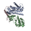
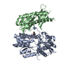
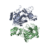
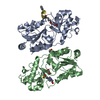
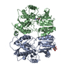
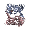
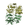
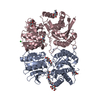
| Title | Crystal structure of human ELOVL fatty acid elongase 7 (ELOVL7) | |||||||||
 Components Components | Elongation of very long chain fatty acids protein 7 | |||||||||
 Keywords Keywords |  TRANSFERASE / Fatty acid biosynthesis / Lipid biosynthesis / membrane protein / Structural Genomics / Structural Genomics Consortium / SGC TRANSFERASE / Fatty acid biosynthesis / Lipid biosynthesis / membrane protein / Structural Genomics / Structural Genomics Consortium / SGC | |||||||||
| Function / homology |  Function and homology informationvery-long-chain 3-oxoacyl-CoA synthase / fatty acid elongase activity / fatty acid elongation, monounsaturated fatty acid / fatty acid elongation, polyunsaturated fatty acid / : / : / : / : / long-chain fatty-acyl-CoA biosynthetic process / Synthesis of very long-chain fatty acyl-CoAs ...very-long-chain 3-oxoacyl-CoA synthase / fatty acid elongase activity / fatty acid elongation, monounsaturated fatty acid / fatty acid elongation, polyunsaturated fatty acid / : / : / : / : / long-chain fatty-acyl-CoA biosynthetic process / Synthesis of very long-chain fatty acyl-CoAs / very long-chain fatty acid biosynthetic process / unsaturated fatty acid biosynthetic process / : / fatty acid elongation, saturated fatty acid / sphingolipid biosynthetic process / endoplasmic reticulum membrane / endoplasmic reticulum Function and homology informationvery-long-chain 3-oxoacyl-CoA synthase / fatty acid elongase activity / fatty acid elongation, monounsaturated fatty acid / fatty acid elongation, polyunsaturated fatty acid / : / : / : / : / long-chain fatty-acyl-CoA biosynthetic process / Synthesis of very long-chain fatty acyl-CoAs ...very-long-chain 3-oxoacyl-CoA synthase / fatty acid elongase activity / fatty acid elongation, monounsaturated fatty acid / fatty acid elongation, polyunsaturated fatty acid / : / : / : / : / long-chain fatty-acyl-CoA biosynthetic process / Synthesis of very long-chain fatty acyl-CoAs / very long-chain fatty acid biosynthetic process / unsaturated fatty acid biosynthetic process / : / fatty acid elongation, saturated fatty acid / sphingolipid biosynthetic process / endoplasmic reticulum membrane / endoplasmic reticulumSimilarity search - Function | |||||||||
| Biological species |  Homo sapiens (human) Homo sapiens (human) | |||||||||
| Method | X-RAY DIFFRACTION / SYNCHROTRON / SIRAS / Resolution: 2.052 Å | |||||||||
 Authors Authors | Nie, L. / Pike, A.C.W. / Bushell, S.R. / Chu, A. / Cole, V. / Speedman, D. / Rodstrom, K.E.J. / Kupinska, K. / Shrestha, L. / Mukhopadhyay, S.M.M. ...Nie, L. / Pike, A.C.W. / Bushell, S.R. / Chu, A. / Cole, V. / Speedman, D. / Rodstrom, K.E.J. / Kupinska, K. / Shrestha, L. / Mukhopadhyay, S.M.M. / Burgess-Brown, N.A. / Love, J. / Edwards, A.M. / Arrowsmith, C.H. / Bountra, C. / Carpenter, E.P. / Structural Genomics Consortium (SGC) | |||||||||
| Funding support |  United Kingdom, 2items United Kingdom, 2items
| |||||||||
 Citation Citation | Journal: TO BE PUBLISHED Title: Crystal structure of human ELOVL fatty acid elongase 7 (ELOVL7) Authors: Nie, L. / Pike, A.C.W. / Bushell, S.R. / Chu, A. / Cole, V. / Speedman, D. / Kupinska, K. / Shrestha, L. / Mukhopadhyay, S.M.M. / Burgess-Brown, N.A. / Love, J. / Edwards, A.M. / Arrowsmith, ...Authors: Nie, L. / Pike, A.C.W. / Bushell, S.R. / Chu, A. / Cole, V. / Speedman, D. / Kupinska, K. / Shrestha, L. / Mukhopadhyay, S.M.M. / Burgess-Brown, N.A. / Love, J. / Edwards, A.M. / Arrowsmith, C.H. / Bountra, C. / Carpenter, E.P. | |||||||||
| History |
|
- Structure visualization
| Structure viewer | Molecule: MolmilJmol/JSmol |
|---|
- Downloads & links
-Download
| PDBx/mmCIF format | 6y7f.cif.gz | 231.5 KB | Display | PDBx/mmCIF format |
|---|---|---|---|---|
| PDB format | pdb6y7f.ent.gz | 197.6 KB | Display | PDB format |
| PDBx/mmJSON format | 6y7f.json.gz | Tree view | PDBx/mmJSON format | |
| Others |  Other downloads Other downloads |
-Validation report
| Arichive directory | https://data.pdbj.org/pub/pdb/validation_reports/y7/6y7fftp://data.pdbj.org/pub/pdb/validation_reports/y7/6y7f | HTTPS FTP |
|---|
-Related structure data
| Similar structure data |
|---|


-Links
 PDBj
PDBj- Assembly
| Deposited unit | 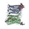
| ||||||||
|---|---|---|---|---|---|---|---|---|---|
| 1 |
| ||||||||
| Unit cell |
|
-Components
| #1: Protein | Mass: 34258.164 Da / Num. of mol.: 2 Source method: isolated from a genetically manipulated source Source: (gene. exp.) Homo sapiens (human) / Gene: ELOVL7 / Plasmid: pFB-CT10HF-LIC / Production host:   Spodoptera frugiperda (fall armyworm) Spodoptera frugiperda (fall armyworm)References: UniProt: A1L3X0, very-long-chain 3-oxoacyl-CoA synthase#2: Chemical |   Mass: 1076.033 Da / Num. of mol.: 2 / Source method: obtained synthetically / Formula: C41H72N7O18P3S / Feature type: SUBJECT OF INVESTIGATION Mass: 1076.033 Da / Num. of mol.: 2 / Source method: obtained synthetically / Formula: C41H72N7O18P3S / Feature type: SUBJECT OF INVESTIGATION#3: Chemical | ChemComp-37X /   Mass: 568.695 Da / Num. of mol.: 4 / Source method: obtained synthetically / Formula: C27H52O12 Mass: 568.695 Da / Num. of mol.: 4 / Source method: obtained synthetically / Formula: C27H52O12#4: Chemical | Chloride  Mass: 35.453 Da / Num. of mol.: 2 / Source method: obtained synthetically / Formula: Cl Mass: 35.453 Da / Num. of mol.: 2 / Source method: obtained synthetically / Formula: Cl#5: Water | ChemComp-HOH / | Water Mass: 18.015 Da / Num. of mol.: 112 / Source method: isolated from a natural source / Formula: H2O Mass: 18.015 Da / Num. of mol.: 112 / Source method: isolated from a natural source / Formula: H2OHas ligand of interest | Y | |
|---|
-Experimental details
-Experiment
| Experiment | Method: X-RAY DIFFRACTION / Number of used crystals: 1 |
|---|
- Sample preparation
| Crystal | Density Matthews: 3.73 Å3/Da / Density % sol: 67.04 % |
|---|---|
| Crystal grow | Temperature: 279 K / Method: vapor diffusion, hanging drop / pH: 4.5 Details: 30-36% PEG400 -- 0.1M acetate pH 4.5 -- 0.23M sodium chloride |
-Data collection
| Diffraction | Mean temperature: 100 K / Serial crystal experiment: N |
|---|---|
| Diffraction source | Source: SYNCHROTRON / Site: Diamond / Beamline: I24 / Wavelength: 0.9686 Å |
| Detector | Type: DECTRIS PILATUS3 6M / Detector: PIXEL / Date: Dec 7, 2019 |
| Radiation | Protocol: SINGLE WAVELENGTH / Monochromatic (M) / Laue (L): M / Scattering type: x-ray |
| Radiation wavelength | Wavelength: 0.9686 Å / Relative weight: 1 |
| Reflection | Resolution: 2.052→62.262 Å / Num. obs: 36724 / % possible obs: 58.7 % / Redundancy: 6.9 % / CC1/2: 0.987 / Rmerge(I) obs: 0.101 / Rpim(I) all: 0.062 / Rrim(I) all: 0.119 / Net I/σ(I): 8.3 |
| Reflection shell | Resolution: 2.052→2.182 Å / Redundancy: 6.2 % / Rmerge(I) obs: 0.703 / Mean I/σ(I) obs: 1.9 / Num. unique obs: 1837 / CC1/2: 0.813 / Rpim(I) all: 0.449 / Rrim(I) all: 0.839 / % possible all: 17.5 |
- Processing
| Software |
| |||||||||||||||||||||||||||||||||||||||||||||||||||||||||||||||||||||||||||
|---|---|---|---|---|---|---|---|---|---|---|---|---|---|---|---|---|---|---|---|---|---|---|---|---|---|---|---|---|---|---|---|---|---|---|---|---|---|---|---|---|---|---|---|---|---|---|---|---|---|---|---|---|---|---|---|---|---|---|---|---|---|---|---|---|---|---|---|---|---|---|---|---|---|---|---|---|
| Refinement | Method to determine structure: SIRAS / Resolution: 2.052→28.68 Å / Cor.coef. Fo:Fc: 0.889 / Cor.coef. Fo:Fc free: 0.876 / SU R Cruickshank DPI: 0.261 / Cross valid method: THROUGHOUT / SU R Blow DPI: 0.251 / SU Rfree Blow DPI: 0.191 / SU Rfree Cruickshank DPI: 0.197 Details: Structure refined against STARANISO anisotropically truncated data. LSSR restraints were used and a single TLS group was refined for each chain. Heterogen dictionaries were generated using ...Details: Structure refined against STARANISO anisotropically truncated data. LSSR restraints were used and a single TLS group was refined for each chain. Heterogen dictionaries were generated using GRADE. ACCOMPANYING SF CIF CONTAINS STARANISO DATA USED FOR REFINEMENT ALONG WITH SCALED MERGED DATA WITHOUT TRUNCATION.
| |||||||||||||||||||||||||||||||||||||||||||||||||||||||||||||||||||||||||||
| Displacement parameters | Biso mean: 43.99 Å2
| |||||||||||||||||||||||||||||||||||||||||||||||||||||||||||||||||||||||||||
| Refine analyze | Luzzati coordinate error obs: 0.29 Å | |||||||||||||||||||||||||||||||||||||||||||||||||||||||||||||||||||||||||||
| Refinement step | Cycle: LAST / Resolution: 2.052→28.68 Å
| |||||||||||||||||||||||||||||||||||||||||||||||||||||||||||||||||||||||||||
| Refine LS restraints |
| |||||||||||||||||||||||||||||||||||||||||||||||||||||||||||||||||||||||||||
| LS refinement shell | Resolution: 2.052→2.1 Å
| |||||||||||||||||||||||||||||||||||||||||||||||||||||||||||||||||||||||||||
| Refinement TLS params. | Refine-ID: X-RAY DIFFRACTION
| |||||||||||||||||||||||||||||||||||||||||||||||||||||||||||||||||||||||||||
| Refinement TLS group |
|
