 Movie
Movie Controller

[English] 日本語
 Yorodumi
- PDB-6y78: Structure of galectin-3C in complex with lactose determined by se... -
+ Open data

- Basic information
| Entry | Database: PDB / ID: 6y78 | ||||||
|---|---|---|---|---|---|---|---|
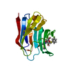
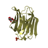
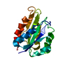
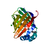
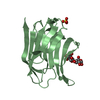
| Title | Structure of galectin-3C in complex with lactose determined by serial crystallography using a silicon nitride membrane support | ||||||
 Components Components | Galectin-3 | ||||||
 Keywords Keywords | SUGAR BINDING PROTEIN / galectin / synchrotron serial crystallography / solid support / silicon nitride membranel | ||||||
| Function / homology |  Function and homology information Function and homology informationnegative regulation of protein tyrosine phosphatase activity / negative regulation of immunological synapse formation / negative regulation of T cell activation via T cell receptor contact with antigen bound to MHC molecule on antigen presenting cell / RUNX2 regulates genes involved in differentiation of myeloid cells / disaccharide binding / regulation of T cell apoptotic process / mononuclear cell migration / IgE binding / negative regulation of endocytosis / positive regulation of mononuclear cell migration ...negative regulation of protein tyrosine phosphatase activity / negative regulation of immunological synapse formation / negative regulation of T cell activation via T cell receptor contact with antigen bound to MHC molecule on antigen presenting cell / RUNX2 regulates genes involved in differentiation of myeloid cells / disaccharide binding / regulation of T cell apoptotic process / mononuclear cell migration / IgE binding / negative regulation of endocytosis / positive regulation of mononuclear cell migration / eosinophil chemotaxis / regulation of extrinsic apoptotic signaling pathway via death domain receptors / RUNX1 regulates transcription of genes involved in differentiation of myeloid cells / protein phosphatase inhibitor activity / negative regulation of T cell receptor signaling pathway / positive chemotaxis / regulation of T cell proliferation / positive regulation of calcium ion import / macrophage chemotaxis / chemoattractant activity / monocyte chemotaxis / Advanced glycosylation endproduct receptor signaling / ficolin-1-rich granule membrane / immunological synapse / laminin binding / epithelial cell differentiation / neutrophil chemotaxis / RNA splicing / secretory granule membrane / molecular condensate scaffold activity / negative regulation of extrinsic apoptotic signaling pathway / positive regulation of protein localization to plasma membrane / spliceosomal complex / positive regulation of protein-containing complex assembly / mRNA processing / carbohydrate binding / protein phosphatase binding / collagen-containing extracellular matrix / mitochondrial inner membrane / innate immune response / Neutrophil degranulation / cell surface / extracellular space / RNA binding / extracellular exosome / extracellular region / nucleoplasm / membrane / nucleus / plasma membrane / cytosol / cytoplasmSimilarity search - Function | ||||||
| Biological species |  Homo sapiens (human) Homo sapiens (human) | ||||||
| Method | X-RAY DIFFRACTION / SYNCHROTRON / MOLECULAR REPLACEMENT / Resolution: 1.7 Å | ||||||
 Authors Authors | Hakansson, M. / Welin, M. / Shilova, A. / Kovacic, R. / Mueller, U. / Logan, D.T. | ||||||
| Funding support |  Sweden, 1items Sweden, 1items
| ||||||
 Citation Citation | Journal: J.Synchrotron Radiat. / Year: 2020 Title: Current status and future opportunities for serial crystallography at MAX IV Laboratory. Authors: Shilova, A. / Lebrette, H. / Aurelius, O. / Nan, J. / Welin, M. / Kovacic, R. / Ghosh, S. / Safari, C. / Friel, R.J. / Milas, M. / Matej, Z. / Hogbom, M. / Branden, G. / Kloos, M. / Shoeman, ...Authors: Shilova, A. / Lebrette, H. / Aurelius, O. / Nan, J. / Welin, M. / Kovacic, R. / Ghosh, S. / Safari, C. / Friel, R.J. / Milas, M. / Matej, Z. / Hogbom, M. / Branden, G. / Kloos, M. / Shoeman, R.L. / Doak, B. / Ursby, T. / Hakansson, M. / Logan, D.T. / Mueller, U. | ||||||
| History |
|
- Structure visualization
| Structure viewer | Molecule: MolmilJmol/JSmol |
|---|
- Downloads & links
-Download
| PDBx/mmCIF format | 6y78.cif.gz | 78.6 KB | Display | PDBx/mmCIF format |
|---|---|---|---|---|
| PDB format | pdb6y78.ent.gz | 55.9 KB | Display | PDB format |
| PDBx/mmJSON format | 6y78.json.gz | Tree view | PDBx/mmJSON format | |
| Others |  Other downloads Other downloads |
-Validation report
| Arichive directory | https://data.pdbj.org/pub/pdb/validation_reports/y7/6y78ftp://data.pdbj.org/pub/pdb/validation_reports/y7/6y78 | HTTPS FTP |
|---|
-Related structure data
| Related structure data | 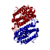 6y2nC  6y4cC  6eymS S: Starting model for refinement C: citing same article ( |
|---|---|
| Similar structure data |


-Links
 PDBj
PDBj


- Assembly
| Deposited unit | 
| ||||||||||
|---|---|---|---|---|---|---|---|---|---|---|---|
| 1 |
| ||||||||||
| Unit cell |
|
-Components
| #1: Protein | / Gal-3 / 35 kDa lectin / Carbohydrate-binding protein 35 / CBP 35 / Galactose-specific lectin 3 / ...Gal-3 / 35 kDa lectin / Carbohydrate-binding protein 35 / CBP 35 / Galactose-specific lectin 3 / Galactoside-binding protein / GALBP / IgE-binding protein / L-31 / Laminin-binding protein / Lectin L-29 / Mac-2 antigen Mass: 15701.049 Da / Num. of mol.: 1 Source method: isolated from a genetically manipulated source Source: (gene. exp.) Homo sapiens (human) / Gene: LGALS3, MAC2 / Production host:  Escherichia coli (E. coli) / References: UniProt: P17931 Escherichia coli (E. coli) / References: UniProt: P17931 |
|---|---|
| #2: Polysaccharide | beta-D-galactopyranose-(1-4)-beta-D-glucopyranose / beta-lactose  , Oligosaccharide / Class: Nutrient / Mass: 342.297 Da / Num. of mol.: 1 , Oligosaccharide / Class: Nutrient / Mass: 342.297 Da / Num. of mol.: 1Source method: isolated from a genetically manipulated source Details: oligosaccharide / References: beta-lactose |
| #3: Water | ChemComp-HOH / Water Mass: 18.015 Da / Num. of mol.: 73 / Source method: isolated from a natural source / Formula: H2O Mass: 18.015 Da / Num. of mol.: 73 / Source method: isolated from a natural source / Formula: H2O |
| Has ligand of interest | Y |
-Experimental details
-Experiment
| Experiment | Method: X-RAY DIFFRACTION / Number of used crystals: 32303 |
|---|
- Sample preparation
| Crystal | Density Matthews: 1.96 Å3/Da / Density % sol: 37.18 % |
|---|---|
| Crystal grow | Temperature: 277 K / Method: vapor diffusion, hanging drop / pH: 7.5 Details: Crystals were grown on XtalTool supports (Jena Bioscience) over a 24 well VDX-plate with 1 ml reservoir. The drop was made up of 1 microlitre 20 mg/ml protein in 10 mM sodium phosphate pH 7. ...Details: Crystals were grown on XtalTool supports (Jena Bioscience) over a 24 well VDX-plate with 1 ml reservoir. The drop was made up of 1 microlitre 20 mg/ml protein in 10 mM sodium phosphate pH 7.4, 100 mM NaCl, 10 mM beta-mercaptoethanol, 2 mM lactose, 0.25 microlitres of galectin-3C seed crystals in a stabilization solution containing 0.1 M Tris pH 7.5, 33% (w/v) PEG 4000, 0.1 M MgCl2, 0.2 M NaSCN and 0.75 microlitres reservoir solution containing 0.1 M Tris pH 7.5, 34% (w/v) PEG 4000, 0.2 M NaSCN and 8 mM beta-mercapthoethanol. Crystals grew to a size of approximately 10-15 micrometer. |
-Data collection
| Diffraction | Mean temperature: 298 K / Serial crystal experiment: Y |
|---|---|
| Diffraction source | Source: SYNCHROTRON / Site: MAX IV / Beamline: BioMAX / Wavelength: 0.98 Å |
| Detector | Type: DECTRIS EIGER X 16M / Detector: PIXEL / Date: Apr 4, 2019 |
| Radiation | Monochromator: DOUBLE CRYSTAL / Protocol: SINGLE WAVELENGTH / Monochromatic (M) / Laue (L): M / Scattering type: x-ray |
| Radiation wavelength | Wavelength: 0.98 Å / Relative weight: 1 |
| Reflection | Resolution: 1.7→61 Å / Num. obs: 14154 / % possible obs: 100 % / Redundancy: 195.2 % / Biso Wilson estimate: 21.27 Å2 / R split: 0.155 / Net I/σ(I): 5.3 |
| Reflection shell | Resolution: 1.7→1.75 Å / Redundancy: 133.1 % / Mean I/σ(I) obs: 1.41 / Num. unique obs: 1369 / R split: 1.833 / % possible all: 100 |
| Serial crystallography measurement | Source size: 100 µm2 |
| Serial crystallography sample delivery | Description: silicon nitride membrane (Silson) / Method: fixed target |
| Serial crystallography sample delivery fixed target | Description: silicon nitride membrane / Motion control: MD3 (Arinax) / Sample dehydration prevention: membrane sandwich / Support base: cryo cap |
| Serial crystallography data reduction | Frame hits: 21029 / Frames failed index: 7507 / Frames indexed: 13522 / Frames total: 32303 |
- Processing
| Software |
| ||||||||||||||||||||||||||||||||||||||||||
|---|---|---|---|---|---|---|---|---|---|---|---|---|---|---|---|---|---|---|---|---|---|---|---|---|---|---|---|---|---|---|---|---|---|---|---|---|---|---|---|---|---|---|---|
| Refinement | Method to determine structure: MOLECULAR REPLACEMENT Starting model: 6EYM Resolution: 1.7→41.45 Å / SU ML: 0.1801 / Cross valid method: FREE R-VALUE / σ(F): 0 / Phase error: 21.9329 Stereochemistry target values: GeoStd + Monomer Library + CDL v1.2
| ||||||||||||||||||||||||||||||||||||||||||
| Solvent computation | Shrinkage radii: 0.9 Å / VDW probe radii: 1.11 Å / Solvent model: FLAT BULK SOLVENT MODEL | ||||||||||||||||||||||||||||||||||||||||||
| Displacement parameters | Biso mean: 28.49 Å2 | ||||||||||||||||||||||||||||||||||||||||||
| Refinement step | Cycle: LAST / Resolution: 1.7→41.45 Å
| ||||||||||||||||||||||||||||||||||||||||||
| Refine LS restraints |
| ||||||||||||||||||||||||||||||||||||||||||
| LS refinement shell |
| ||||||||||||||||||||||||||||||||||||||||||
| Refinement TLS params. | Method: refined / Origin x: 12.6737030943 Å / Origin y: 0.42243450904 Å / Origin z: 6.2913268179 Å
| ||||||||||||||||||||||||||||||||||||||||||
| Refinement TLS group | Selection details: all |
