 Movie
Movie Controller

[English] 日本語
 Yorodumi
- PDB-6y4b: Structure of cyclodipeptide synthase from Candidatus Glomeribacte... -
+ Open data

- Basic information
| Entry | Database: PDB / ID: 6y4b | ||||||
|---|---|---|---|---|---|---|---|
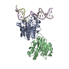
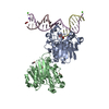
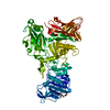
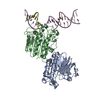
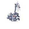
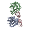
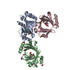
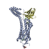
| Title | Structure of cyclodipeptide synthase from Candidatus Glomeribacter gigasporarum bound to Phe-tRNAPhe | ||||||
 Components Components |
| ||||||
 Keywords Keywords |  RNA BINDING PROTEIN / cyclodipeptide synthase / tRNA / Complex RNA BINDING PROTEIN / cyclodipeptide synthase / tRNA / Complex | ||||||
| Function / homology | PHENYLALANINE / RNA / RNA (> 10) / Uncharacterized protein Function and homology information Function and homology information | ||||||
| Biological species |  Escherichia coli (E. coli)Candidatus Glomeribacter gigasporarum BEG34 (bacteria) Escherichia coli (E. coli)Candidatus Glomeribacter gigasporarum BEG34 (bacteria) | ||||||
| Method | X-RAY DIFFRACTION / SYNCHROTRON / MOLECULAR REPLACEMENT / Resolution: 5 Å | ||||||
 Authors Authors | Bourgeois, G. / Mechulam, Y. / Schmitt, E. | ||||||
| Funding support |  France, 1items France, 1items
| ||||||
 Citation Citation | Journal: Rna / Year: 2020 Title: Structural basis of the interaction between cyclodipeptide synthases and aminoacylated tRNA substrates. Authors: Bourgeois, G. / Seguin, J. / Babin, M. / Gondry, M. / Mechulam, Y. / Schmitt, E. | ||||||
| History |
|
- Structure visualization
| Structure viewer | Molecule: MolmilJmol/JSmol |
|---|
- Downloads & links
-Download
| PDBx/mmCIF format | 6y4b.cif.gz | 207.1 KB | Display | PDBx/mmCIF format |
|---|---|---|---|---|
| PDB format | pdb6y4b.ent.gz | 135 KB | Display | PDB format |
| PDBx/mmJSON format | 6y4b.json.gz | Tree view | PDBx/mmJSON format | |
| Others |  Other downloads Other downloads |
-Validation report
| Arichive directory | https://data.pdbj.org/pub/pdb/validation_reports/y4/6y4bftp://data.pdbj.org/pub/pdb/validation_reports/y4/6y4b | HTTPS FTP |
|---|
-Related structure data
| Related structure data | 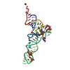 6y3gC 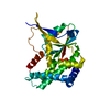 5mlpS S: Starting model for refinement C: citing same article ( |
|---|---|
| Similar structure data |


-Links
 PDBj
PDBj


- Assembly
| Deposited unit | 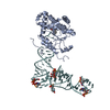
| ||||||||||||
|---|---|---|---|---|---|---|---|---|---|---|---|---|---|
| 1 |
| ||||||||||||
| Unit cell |
|
-Components
| #1: RNA chain | Mass: 24519.662 Da / Num. of mol.: 1 Source method: isolated from a genetically manipulated source Details: tRNA is aminoacylated, adenosine 76 is bounded to phenylalanine Source: (gene. exp.) Escherichia coli (E. coli) / Production host: Escherichia coli (E. coli) |
|---|---|
| #2: Protein | Mass: 34119.309 Da / Num. of mol.: 1 Source method: isolated from a genetically manipulated source Source: (gene. exp.) Candidatus Glomeribacter gigasporarum BEG34 (bacteria)Gene: CAGGBEG34_30028 / Production host: Escherichia coli (E. coli) / References: UniProt: G2JBB2 |
| #3: Chemical | ChemComp-PHE / Phenylalanine  Type: L-peptide linking / Mass: 165.189 Da / Num. of mol.: 1 / Source method: obtained synthetically / Formula: C9H11NO2 Type: L-peptide linking / Mass: 165.189 Da / Num. of mol.: 1 / Source method: obtained synthetically / Formula: C9H11NO2 |
| Has ligand of interest | N |
-Experimental details
-Experiment
| Experiment | Method: X-RAY DIFFRACTION / Number of used crystals: 1 |
|---|
- Sample preparation
| Crystal | Density Matthews: 5.55 Å3/Da / Density % sol: 77.84 % |
|---|---|
| Crystal grow | Temperature: 278 K / Method: vapor diffusion, hanging drop Details: 10% PEG8000, 0.17 M AcCa, 0.17 M Guanidinium hydrochloride, 0.1 M Hepes pH 7 |
-Data collection
| Diffraction | Mean temperature: 100 K / Serial crystal experiment: N |
|---|---|
| Diffraction source | Source: SYNCHROTRON / Site: SOLEIL / Beamline: PROXIMA 2 / Wavelength: 0.9786 Å |
| Detector | Type: DECTRIS PILATUS 6M / Detector: PIXEL / Date: Sep 16, 2016 |
| Radiation | Protocol: SINGLE WAVELENGTH / Monochromatic (M) / Laue (L): M / Scattering type: x-ray |
| Radiation wavelength | Wavelength: 0.9786 Å / Relative weight: 1 |
| Reflection | Resolution: 5→50 Å / Num. obs: 6120 / % possible obs: 95.6 % / Redundancy: 33.2 % / Biso Wilson estimate: 214.56 Å2 / CC1/2: 0.99 / Net I/σ(I): 10.7 |
| Reflection shell | Resolution: 5→5.3 Å / Redundancy: 32 % / Mean I/σ(I) obs: 1 / Num. unique obs: 867 / CC1/2: 0.58 / % possible all: 91.6 |
- Processing
| Software |
| ||||||||||||||||||||||||||||||||||||||||
|---|---|---|---|---|---|---|---|---|---|---|---|---|---|---|---|---|---|---|---|---|---|---|---|---|---|---|---|---|---|---|---|---|---|---|---|---|---|---|---|---|---|
| Refinement | Method to determine structure: MOLECULAR REPLACEMENT Starting model: 5MLP Resolution: 5→48.16 Å / SU ML: 1.0625 / Cross valid method: FREE R-VALUE / σ(F): 1.33 / Phase error: 36.2752 Stereochemistry target values: GeoStd + Monomer Library + CDL v1.2
| ||||||||||||||||||||||||||||||||||||||||
| Solvent computation | Shrinkage radii: 0.9 Å / VDW probe radii: 1.11 Å / Solvent model: FLAT BULK SOLVENT MODEL | ||||||||||||||||||||||||||||||||||||||||
| Displacement parameters | Biso mean: 139.07 Å2 | ||||||||||||||||||||||||||||||||||||||||
| Refinement step | Cycle: LAST / Resolution: 5→48.16 Å
| ||||||||||||||||||||||||||||||||||||||||
| Refine LS restraints |
| ||||||||||||||||||||||||||||||||||||||||
| LS refinement shell |
| ||||||||||||||||||||||||||||||||||||||||
| Refinement TLS params. | Method: refined / Origin x: -80.2336000483 Å / Origin y: 69.1408391067 Å / Origin z: 3.14746588878 Å
| ||||||||||||||||||||||||||||||||||||||||
| Refinement TLS group | Selection details: all |
