 Movie
Movie Controller

[English] 日本語
 Yorodumi
- PDB-6xiz: Crystal structure of multi-copper oxidase from Pediococcus acidil... -
+ Open data

- Basic information
| Entry | Database: PDB / ID: 6xiz | |||||||||||||||||||||
|---|---|---|---|---|---|---|---|---|---|---|---|---|---|---|---|---|---|---|---|---|---|---|
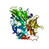
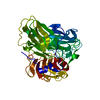
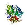
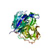
| Title | Crystal structure of multi-copper oxidase from Pediococcus acidilactici | |||||||||||||||||||||
 Components Components | Copper oxidase | |||||||||||||||||||||
 Keywords Keywords |  OXIDOREDUCTASE / LACCASE / COPPER OXIDASE / ACOUSTIC DROPLET EJECTION / LIGNIN / COMBINATORIAL CRYSTALLIZATION OXIDOREDUCTASE / LACCASE / COPPER OXIDASE / ACOUSTIC DROPLET EJECTION / LIGNIN / COMBINATORIAL CRYSTALLIZATION | |||||||||||||||||||||
| Function / homology |  Function and homology information Function and homology information | |||||||||||||||||||||
| Biological species |  Pediococcus acidilactici (bacteria) Pediococcus acidilactici (bacteria) | |||||||||||||||||||||
| Method | X-RAY DIFFRACTION / SYNCHROTRON / SAD / Resolution: 1.8 Å | |||||||||||||||||||||
 Authors Authors | Pardo, I. / Soares, A.S. / Collins, R. / Partowmah, S.H. / Coler, E.A. | |||||||||||||||||||||
| Funding support |  United States, 6items United States, 6items
| |||||||||||||||||||||
 Citation Citation | Journal: Microb Biotechnol / Year: 2021 Title: Structural analysis and biochemical properties of laccase enzymes from two Pediococcus species. Authors: Olmeda, I. / Casino, P. / Collins, R.E. / Sendra, R. / Callejon, S. / Huesa, J. / Soares, A.S. / Ferrer, S. / Pardo, I. | |||||||||||||||||||||
| History |
|
- Structure visualization
| Structure viewer | Molecule: MolmilJmol/JSmol |
|---|
- Downloads & links
-Download
| PDBx/mmCIF format | 6xiz.cif.gz | 425.5 KB | Display | PDBx/mmCIF format |
|---|---|---|---|---|
| PDB format | pdb6xiz.ent.gz | 347.9 KB | Display | PDB format |
| PDBx/mmJSON format | 6xiz.json.gz | Tree view | PDBx/mmJSON format | |
| Others |  Other downloads Other downloads |
-Validation report
| Arichive directory | https://data.pdbj.org/pub/pdb/validation_reports/xi/6xizftp://data.pdbj.org/pub/pdb/validation_reports/xi/6xiz | HTTPS FTP |
|---|
-Related structure data


-Links
 PDBj
PDBj
- Assembly
| Deposited unit | 
| ||||||||
|---|---|---|---|---|---|---|---|---|---|
| 1 | 
| ||||||||
| 2 | 
| ||||||||
| Unit cell |
|
-Components
-Protein , 1 types, 2 molecules AB
| #1: Protein | Mass: 54385.145 Da / Num. of mol.: 2 Source method: isolated from a genetically manipulated source Source: (gene. exp.) Pediococcus acidilactici (bacteria) / Gene: BTW26_06860, FEZ49_06610 / Production host: Escherichia coli (E. coli) / References: UniProt: A0A1A5VCP7 |
|---|
-Non-polymers , 5 types, 618 molecules 


| #2: Chemical | ChemComp-CU / Copper Mass: 63.546 Da / Num. of mol.: 4 / Source method: obtained synthetically / Formula: Cu / Feature type: SUBJECT OF INVESTIGATION Mass: 63.546 Da / Num. of mol.: 4 / Source method: obtained synthetically / Formula: Cu / Feature type: SUBJECT OF INVESTIGATION#3: Chemical | Copper(I) oxide Mass: 143.091 Da / Num. of mol.: 2 / Source method: obtained synthetically / Formula: Cu2O / Feature type: SUBJECT OF INVESTIGATION Mass: 143.091 Da / Num. of mol.: 2 / Source method: obtained synthetically / Formula: Cu2O / Feature type: SUBJECT OF INVESTIGATION#4: Chemical | Chloride Mass: 35.453 Da / Num. of mol.: 2 / Source method: obtained synthetically / Formula: Cl Mass: 35.453 Da / Num. of mol.: 2 / Source method: obtained synthetically / Formula: Cl#5: Chemical | ChemComp-BEN / | Benzamidine Mass: 120.152 Da / Num. of mol.: 1 / Source method: obtained synthetically / Formula: C7H8N2 Mass: 120.152 Da / Num. of mol.: 1 / Source method: obtained synthetically / Formula: C7H8N2#6: Water | ChemComp-HOH / | WaterMass: 18.015 Da / Num. of mol.: 609 / Source method: isolated from a natural source / Formula: H2O |
|---|
-Details
| Has ligand of interest | Y |
|---|
-Experimental details
-Experiment
| Experiment | Method: X-RAY DIFFRACTION / Number of used crystals: 1 |
|---|
- Sample preparation
| Crystal | Density Matthews: 2.47 Å3/Da / Density % sol: 50.22 % |
|---|---|
| Crystal grow | Temperature: 292 K / Method: vapor diffusion, sitting drop Details: 52.5 nL drop composed of 95 mM Sodium Citrate buffer, pH 3.0, 3% polyethylene glycol 8000, 7.8% polyethylene glycol 400, 1.2% dimethyl sulfoxide, 114 mM benzamidine hydrochloride, and 5.0-10. ...Details: 52.5 nL drop composed of 95 mM Sodium Citrate buffer, pH 3.0, 3% polyethylene glycol 8000, 7.8% polyethylene glycol 400, 1.2% dimethyl sulfoxide, 114 mM benzamidine hydrochloride, and 5.0-10.0 mg/mL protein in 5 mM Tris-HCL pH 7.2 Mylar was initially used as the surface, resulting in thin polycrystalline needles. Changing the surface to COC (cyclic olefin copolymer) resulted in large high-quality single crystals. |
-Data collection
| Diffraction | Mean temperature: 100 K / Serial crystal experiment: N |
|---|---|
| Diffraction source | Source: SYNCHROTRON / Site: NSLS-II / Beamline: 17-ID-1 / Wavelength: 0.9202 Å |
| Detector | Type: DECTRIS EIGER X 9M / Detector: PIXEL / Date: Aug 10, 2019 |
| Radiation | Protocol: SINGLE WAVELENGTH / Monochromatic (M) / Laue (L): M / Scattering type: x-ray |
| Radiation wavelength | Wavelength: 0.9202 Å / Relative weight: 1 |
| Reflection | Resolution: 1.78→73.65 Å / Num. obs: 98773 / % possible obs: 98.3 % / Redundancy: 25.53 % / CC1/2: 0.999 / Rmerge(I) obs: 0.185 / Net I/σ(I): 10.37 |
| Reflection shell | Resolution: 1.78→1.82 Å / Rmerge(I) obs: 0.4263 / Mean I/σ(I) obs: 0.71 / Num. unique obs: 5726 / CC1/2: 0.476 / % possible all: 77.6 |
- Processing
| Software |
| ||||||||||||||||||||||||||||||||||||||||||||||||||||||||||||||||||||||||||||||||||||||||||||||||||||||||||||||||||||||||||||||||||||||||||||||||||||||||||||||||||||||||||||||||||||||
|---|---|---|---|---|---|---|---|---|---|---|---|---|---|---|---|---|---|---|---|---|---|---|---|---|---|---|---|---|---|---|---|---|---|---|---|---|---|---|---|---|---|---|---|---|---|---|---|---|---|---|---|---|---|---|---|---|---|---|---|---|---|---|---|---|---|---|---|---|---|---|---|---|---|---|---|---|---|---|---|---|---|---|---|---|---|---|---|---|---|---|---|---|---|---|---|---|---|---|---|---|---|---|---|---|---|---|---|---|---|---|---|---|---|---|---|---|---|---|---|---|---|---|---|---|---|---|---|---|---|---|---|---|---|---|---|---|---|---|---|---|---|---|---|---|---|---|---|---|---|---|---|---|---|---|---|---|---|---|---|---|---|---|---|---|---|---|---|---|---|---|---|---|---|---|---|---|---|---|---|---|---|---|---|
| Refinement | Method to determine structure: SAD / Resolution: 1.8→73.65 Å / Cor.coef. Fo:Fc: 0.988 / Cor.coef. Fo:Fc free: 0.98 / SU B: 6.337 / SU ML: 0.076 / Cross valid method: THROUGHOUT / ESU R: 0.144 / ESU R Free: 0.089 / Stereochemistry target values: MAXIMUM LIKELIHOOD / Details: HYDROGENS HAVE BEEN ADDED IN THE RIDING POSITIONS
| ||||||||||||||||||||||||||||||||||||||||||||||||||||||||||||||||||||||||||||||||||||||||||||||||||||||||||||||||||||||||||||||||||||||||||||||||||||||||||||||||||||||||||||||||||||||
| Solvent computation | Ion probe radii: 0.8 Å / Shrinkage radii: 0.8 Å / VDW probe radii: 1.2 Å / Solvent model: MASK | ||||||||||||||||||||||||||||||||||||||||||||||||||||||||||||||||||||||||||||||||||||||||||||||||||||||||||||||||||||||||||||||||||||||||||||||||||||||||||||||||||||||||||||||||||||||
| Displacement parameters | Biso mean: 47.028 Å2
| ||||||||||||||||||||||||||||||||||||||||||||||||||||||||||||||||||||||||||||||||||||||||||||||||||||||||||||||||||||||||||||||||||||||||||||||||||||||||||||||||||||||||||||||||||||||
| Refinement step | Cycle: 1 / Resolution: 1.8→73.65 Å
| ||||||||||||||||||||||||||||||||||||||||||||||||||||||||||||||||||||||||||||||||||||||||||||||||||||||||||||||||||||||||||||||||||||||||||||||||||||||||||||||||||||||||||||||||||||||
| Refine LS restraints |
|