 Movie
Movie Controller

[English] 日本語
 Yorodumi
- PDB-6xh0: Co-crystal structure of HIV-1 TAR RNA in complex with lab-evolved... -
+ Open data

- Basic information
| Entry | Database: PDB / ID: 6xh0 | ||||||
|---|---|---|---|---|---|---|---|
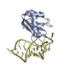
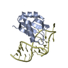
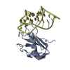
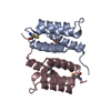
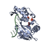
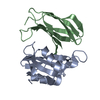
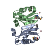
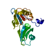
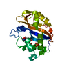
| Title | Co-crystal structure of HIV-1 TAR RNA in complex with lab-evolved RRM TBP6.9 | ||||||
 Components Components |
| ||||||
 Keywords Keywords | RNA BINDING PROTEIN/RNA / PROTEIN-RNA COMPLEX / TAR RNA / LAB-EVOLVED PROTEIN / ARGININE FORK /  BETA HAIRPIN / MAJOR-GROOVE READOUT / BASE TRIPLE / U1A / HIV-1 / TRANS- ACTIVATION / RNA RECOGNITION MOTIF / RRM / RNA BINDING PROTEIN-RNA COMPLEX / RNA BINDING PROTEIN BETA HAIRPIN / MAJOR-GROOVE READOUT / BASE TRIPLE / U1A / HIV-1 / TRANS- ACTIVATION / RNA RECOGNITION MOTIF / RRM / RNA BINDING PROTEIN-RNA COMPLEX / RNA BINDING PROTEIN | ||||||
| Function / homology |  Function and homology information Function and homology informationU1 snRNP binding / U1 snRNP / U1 snRNA binding / U4/U6 x U5 tri-snRNP complex / mRNA Splicing - Major Pathway / spliceosomal complex / mRNA splicing, via spliceosome / DNA binding / RNA binding / nucleoplasm ...U1 snRNP binding / U1 snRNP / U1 snRNA binding / U4/U6 x U5 tri-snRNP complex / mRNA Splicing - Major Pathway / spliceosomal complex / mRNA splicing, via spliceosome / DNA binding / RNA binding / nucleoplasm / identical protein binding / nucleusSimilarity search - Function | ||||||
| Biological species |  Homo sapiens (human) Homo sapiens (human)  Human immunodeficiency virus 1 Human immunodeficiency virus 1 | ||||||
| Method | X-RAY DIFFRACTION / SYNCHROTRON / FOURIER SYNTHESIS / Resolution: 3.1 Å | ||||||
 Authors Authors | Chavali, S.S. / Jenkins, J.L. / Wedekind, J.E. | ||||||
| Funding support |  United States, 1items United States, 1items
| ||||||
 Citation Citation | Journal: J.Biol.Chem. / Year: 2020 Title: Co-crystal structures of HIV TAR RNA bound to lab-evolved proteins show key roles for arginine relevant to the design of cyclic peptide TAR inhibitors. Authors: Chavali, S.S. / Mali, S.M. / Jenkins, J.L. / Fasan, R. / Wedekind, J.E. | ||||||
| History |
|
- Structure visualization
| Structure viewer | Molecule: MolmilJmol/JSmol |
|---|
- Downloads & links
-Download
| PDBx/mmCIF format | 6xh0.cif.gz | 102.6 KB | Display | PDBx/mmCIF format |
|---|---|---|---|---|
| PDB format | pdb6xh0.ent.gz | 77.6 KB | Display | PDB format |
| PDBx/mmJSON format | 6xh0.json.gz | Tree view | PDBx/mmJSON format | |
| Others |  Other downloads Other downloads |
-Validation report
| Arichive directory | https://data.pdbj.org/pub/pdb/validation_reports/xh/6xh0ftp://data.pdbj.org/pub/pdb/validation_reports/xh/6xh0 | HTTPS FTP |
|---|
-Related structure data


-Links
 PDBj
PDBj


- Assembly
| Deposited unit | 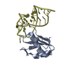
| ||||||||
|---|---|---|---|---|---|---|---|---|---|
| 1 |
| ||||||||
| Unit cell |
|
-Components
| #1: Protein | Mass: 10071.694 Da / Num. of mol.: 1 Source method: isolated from a genetically manipulated source Source: (gene. exp.) Homo sapiens (human) / Production host:  Escherichia coli (E. coli) / References: UniProt: P09012*PLUS Escherichia coli (E. coli) / References: UniProt: P09012*PLUS | ||||
|---|---|---|---|---|---|
| #2: RNA chain | Mass: 8657.167 Da / Num. of mol.: 1 / Source method: obtained synthetically / Source: (synth.) Human immunodeficiency virus 1 | ||||
| #3: Chemical |   Mass: 24.305 Da / Num. of mol.: 2 / Source method: isolated from a natural source / Formula: Mg Mass: 24.305 Da / Num. of mol.: 2 / Source method: isolated from a natural source / Formula: Mg#4: Water | ChemComp-HOH / | Water Mass: 18.015 Da / Num. of mol.: 3 / Source method: isolated from a natural source / Formula: H2O Mass: 18.015 Da / Num. of mol.: 3 / Source method: isolated from a natural source / Formula: H2OHas ligand of interest | N | |
-Experimental details
-Experiment
| Experiment | Method: X-RAY DIFFRACTION / Number of used crystals: 1 |
|---|
- Sample preparation
| Crystal | Density Matthews: 3.13 Å3/Da / Density % sol: 60.76 % |
|---|---|
| Crystal grow | Temperature: 293 K / Method: vapor diffusion, hanging drop / pH: 5.6 Details: 17.5% PEG 4000, 0.05 M MES Monohydrate pH 6.0, 0.005 M MgCl2(H20)6 |
-Data collection
| Diffraction | Mean temperature: 100 K / Serial crystal experiment: N |
|---|---|
| Diffraction source | Source: SYNCHROTRON / Site: SSRL / Beamline: BL12-2 / Wavelength: 0.9795 Å |
| Detector | Type: DECTRIS PILATUS 6M / Detector: PIXEL / Date: Jun 8, 2018 Details: Rh coated collimating mirrors, K-B focusing mirrors |
| Radiation | Monochromator: LIQUID NITROGEN-COOLED DOUBLE CRYSTAL SI(111) Protocol: SINGLE WAVELENGTH / Monochromatic (M) / Laue (L): M / Scattering type: x-ray |
| Radiation wavelength | Wavelength: 0.9795 Å / Relative weight: 1 |
| Reflection | Resolution: 3.1→38.97 Å / Num. obs: 4920 / % possible obs: 99.7 % / Redundancy: 11.5 % / CC1/2: 0.998 / Rpim(I) all: 0.071 / Net I/σ(I): 8.2 |
| Reflection shell | Resolution: 3.1→3.31 Å / Redundancy: 12.1 % / Mean I/σ(I) obs: 1.6 / Num. unique obs: 858 / CC1/2: 0.743 / Rpim(I) all: 0.447 / % possible all: 100 |
- Processing
| Software |
| |||||||||||||||||||||||||||||||||||||||||||||||||||||||||||||||||||||||||||
|---|---|---|---|---|---|---|---|---|---|---|---|---|---|---|---|---|---|---|---|---|---|---|---|---|---|---|---|---|---|---|---|---|---|---|---|---|---|---|---|---|---|---|---|---|---|---|---|---|---|---|---|---|---|---|---|---|---|---|---|---|---|---|---|---|---|---|---|---|---|---|---|---|---|---|---|---|
| Refinement | Method to determine structure: FOURIER SYNTHESIS / Resolution: 3.1→38.966 Å / SU ML: 0.51 / Cross valid method: THROUGHOUT / σ(F): 1.35 / Phase error: 26.52 / Stereochemistry target values: ML
| |||||||||||||||||||||||||||||||||||||||||||||||||||||||||||||||||||||||||||
| Solvent computation | Shrinkage radii: 0.9 Å / VDW probe radii: 1.11 Å / Solvent model: FLAT BULK SOLVENT MODEL | |||||||||||||||||||||||||||||||||||||||||||||||||||||||||||||||||||||||||||
| Displacement parameters | Biso max: 211.89 Å2 / Biso mean: 83.3428 Å2 / Biso min: 56.2 Å2 | |||||||||||||||||||||||||||||||||||||||||||||||||||||||||||||||||||||||||||
| Refinement step | Cycle: final / Resolution: 3.1→38.966 Å
| |||||||||||||||||||||||||||||||||||||||||||||||||||||||||||||||||||||||||||
| LS refinement shell | Refine-ID: X-RAY DIFFRACTION / Rfactor Rfree error: 0
| |||||||||||||||||||||||||||||||||||||||||||||||||||||||||||||||||||||||||||
| Refinement TLS params. | Method: refined / Refine-ID: X-RAY DIFFRACTION
| |||||||||||||||||||||||||||||||||||||||||||||||||||||||||||||||||||||||||||
| Refinement TLS group |
|
