 Movie
Movie Controller


+ Open data

- Basic information
| Entry | Database: PDB / ID: 6xfj | |||||||||
|---|---|---|---|---|---|---|---|---|---|---|
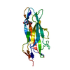
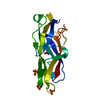
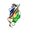
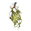
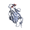
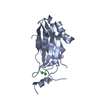
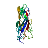
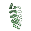
| Title | Crystal structure of the type III secretion pilotin InvH | |||||||||
 Components Components | Type 3 secretion system pilotin Type three secretion system Type three secretion system | |||||||||
 Keywords Keywords | PROTEIN TRANSPORT / pilotin / chaperone / type III secretion / dimer | |||||||||
| Function / homology | InvH outer membrane lipoprotein / InvH outer membrane lipoprotein / cell outer membrane / Prokaryotic membrane lipoprotein lipid attachment site profile. / : / SPI-1 type 3 secretion system pilotin Function and homology information Function and homology information | |||||||||
| Biological species |  Salmonella typhimurium (bacteria) Salmonella typhimurium (bacteria) | |||||||||
| Method | X-RAY DIFFRACTION / SYNCHROTRON / SAD / Resolution: 1.2 Å | |||||||||
 Authors Authors | Majewski, D.D. / Okon, M. / Heinkel, F. / Robb, C.S. / Vuckovic, M. / McIntosh, L.P. / Strynadka, N.C.J. | |||||||||
| Funding support |  Canada, 2items Canada, 2items
| |||||||||
 Citation Citation | Journal: Structure / Year: 2021 Title: Characterization of the Pilotin-Secretin Complex from the Salmonella enterica Type III Secretion System Using Hybrid Structural Methods. Authors: Majewski, D.D. / Okon, M. / Heinkel, F. / Robb, C.S. / Vuckovic, M. / McIntosh, L.P. / Strynadka, N.C.J. | |||||||||
| History |
|
- Structure visualization
| Structure viewer | Molecule: MolmilJmol/JSmol |
|---|
- Downloads & links
-Download
| PDBx/mmCIF format | 6xfj.cif.gz | 164.1 KB | Display | PDBx/mmCIF format |
|---|---|---|---|---|
| PDB format | pdb6xfj.ent.gz | 130.5 KB | Display | PDB format |
| PDBx/mmJSON format | 6xfj.json.gz | Tree view | PDBx/mmJSON format | |
| Others |  Other downloads Other downloads |
-Validation report
| Arichive directory | https://data.pdbj.org/pub/pdb/validation_reports/xf/6xfjftp://data.pdbj.org/pub/pdb/validation_reports/xf/6xfj | HTTPS FTP |
|---|
-Related structure data


-Links
 PDBj
PDBj


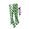
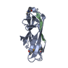
- Assembly
| Deposited unit | 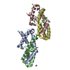
| ||||||||
|---|---|---|---|---|---|---|---|---|---|
| 1 | 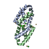
| ||||||||
| 2 | 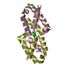
| ||||||||
| Unit cell |
|
-Components
| #1: Protein | Type three secretion system / Invasion lipoprotein invH Mass: 9453.652 Da / Num. of mol.: 4 Source method: isolated from a genetically manipulated source Source: (gene. exp.) Salmonella typhimurium (strain LT2 / SGSC1412 / ATCC 700720) (bacteria)Strain: LT2 / SGSC1412 / ATCC 700720 / Gene: invH, sctG, STM2900 / Production host: Escherichia coli BL21(DE3) (bacteria) / References: UniProt: P0CL43#2: Chemical | ChemComp-CD /   Mass: 112.411 Da / Num. of mol.: 16 / Source method: obtained synthetically / Formula: Cd Mass: 112.411 Da / Num. of mol.: 16 / Source method: obtained synthetically / Formula: Cd#3: Chemical | ChemComp-CL / Chloride  Mass: 35.453 Da / Num. of mol.: 18 / Source method: obtained synthetically / Formula: Cl Mass: 35.453 Da / Num. of mol.: 18 / Source method: obtained synthetically / Formula: Cl#4: Chemical | ChemComp-NA / |   Mass: 22.990 Da / Num. of mol.: 1 / Source method: isolated from a natural source / Formula: Na Mass: 22.990 Da / Num. of mol.: 1 / Source method: isolated from a natural source / Formula: Na#5: Water | ChemComp-HOH / | Water Mass: 18.015 Da / Num. of mol.: 405 / Source method: isolated from a natural source / Formula: H2O Mass: 18.015 Da / Num. of mol.: 405 / Source method: isolated from a natural source / Formula: H2OHas ligand of interest | N | |
|---|
-Experimental details
-Experiment
| Experiment | Method: X-RAY DIFFRACTION / Number of used crystals: 1 |
|---|
- Sample preparation
| Crystal | Density Matthews: 1.91 Å3/Da / Density % sol: 35.45 % |
|---|---|
| Crystal grow | Temperature: 303 K / Method: microbatch / pH: 4.6 Details: 80 mM cadmium chloride, 20% PEG 300, 400 mM NaCl, 100 mM sodium acetate |
-Data collection
| Diffraction | Mean temperature: 100 K / Serial crystal experiment: N |
|---|---|
| Diffraction source | Source: SYNCHROTRON / Site: CLSI / Beamline: 08ID-1 / Wavelength: 0.98 Å |
| Detector | Type: RAYONIX MX-300 / Detector: CCD / Date: Mar 9, 2016 |
| Radiation | Protocol: SINGLE WAVELENGTH / Monochromatic (M) / Laue (L): M / Scattering type: x-ray |
| Radiation wavelength | Wavelength: 0.98 Å / Relative weight: 1 |
| Reflection | Resolution: 1.2→44.36 Å / Num. obs: 82540 / % possible obs: 94.5 % / Redundancy: 3.9 % / CC1/2: 0.998 / Net I/σ(I): 11.1 |
| Reflection shell | Resolution: 1.2→1.24 Å / Num. unique obs: 7937 / CC1/2: 0.681 |
- Processing
| Software |
| |||||||||||||||||||||||||||||||||||||||||||||||||||||||||||||||||
|---|---|---|---|---|---|---|---|---|---|---|---|---|---|---|---|---|---|---|---|---|---|---|---|---|---|---|---|---|---|---|---|---|---|---|---|---|---|---|---|---|---|---|---|---|---|---|---|---|---|---|---|---|---|---|---|---|---|---|---|---|---|---|---|---|---|---|
| Refinement | Method to determine structure: SAD / Resolution: 1.2→44.36 Å / Cor.coef. Fo:Fc: 0.978 / Cor.coef. Fo:Fc free: 0.965 / SU B: 1.969 / SU ML: 0.037 / Cross valid method: THROUGHOUT / σ(F): 0 / ESU R: 0.04 / ESU R Free: 0.041 / Stereochemistry target values: MAXIMUM LIKELIHOOD Details: HYDROGENS HAVE BEEN ADDED IN THE RIDING POSITIONS U VALUES : REFINED INDIVIDUALLY
| |||||||||||||||||||||||||||||||||||||||||||||||||||||||||||||||||
| Solvent computation | Ion probe radii: 0.8 Å / Shrinkage radii: 0.8 Å / VDW probe radii: 1.2 Å / Solvent model: MASK | |||||||||||||||||||||||||||||||||||||||||||||||||||||||||||||||||
| Displacement parameters | Biso max: 77.77 Å2 / Biso mean: 14.865 Å2 / Biso min: 7.87 Å2
| |||||||||||||||||||||||||||||||||||||||||||||||||||||||||||||||||
| Refinement step | Cycle: final / Resolution: 1.2→44.36 Å
| |||||||||||||||||||||||||||||||||||||||||||||||||||||||||||||||||
| Refine LS restraints |
| |||||||||||||||||||||||||||||||||||||||||||||||||||||||||||||||||
| LS refinement shell | Resolution: 1.2→1.231 Å / Rfactor Rfree error: 0 / Total num. of bins used: 20
|
