 Movie
Movie Controller


+ Open data

- Basic information
| Entry | Database: PDB / ID: 6xdj | ||||||
|---|---|---|---|---|---|---|---|
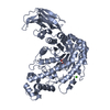
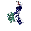
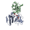
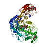
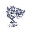
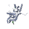
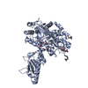
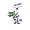
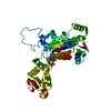
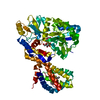
| Title | Crystal Structure Analysis of MBP-SIN3 | ||||||
 Components Components | Maltodextrin-binding protein,Transcriptional regulatory protein SIN3 | ||||||
 Keywords Keywords |  TRANSCRIPTION / RPD3 histone deacetylase complexes / epigenetic repression TRANSCRIPTION / RPD3 histone deacetylase complexes / epigenetic repression | ||||||
| Function / homology |  Function and homology information Function and homology informationnegative regulation of silent mating-type cassette heterochromatin formation / Rpd3L complex / Rpd3S complex / Rpd3L-Expanded complex / negative regulation of rDNA heterochromatin formation / rDNA chromatin condensation / nucleophagy / regulation of DNA-templated DNA replication initiation / negative regulation of transcription by RNA polymerase I / Sin3-type complex ...negative regulation of silent mating-type cassette heterochromatin formation / Rpd3L complex / Rpd3S complex / Rpd3L-Expanded complex / negative regulation of rDNA heterochromatin formation / rDNA chromatin condensation / nucleophagy / regulation of DNA-templated DNA replication initiation / negative regulation of transcription by RNA polymerase I / Sin3-type complex / carbohydrate transmembrane transporter activity / nucleosome assembly / double-strand break repair via nonhomologous end joining / transcription corepressor activity / transcription coactivator activity / periplasmic space / cell cycle / cell division / chromatin / regulation of transcription by RNA polymerase II / negative regulation of transcription by RNA polymerase II / positive regulation of transcription by RNA polymerase II / identical protein binding / nucleusSimilarity search - Function | ||||||
| Biological species |  Serratia sp. (bacteria) Serratia sp. (bacteria) Saccharomyces cerevisiae (brewer's yeast) Saccharomyces cerevisiae (brewer's yeast) | ||||||
| Method | X-RAY DIFFRACTION / SYNCHROTRON / MOLECULAR REPLACEMENT / molecular replacement / Resolution: 2.2 Å | ||||||
 Authors Authors | Seo, H.-S. / Dhe-Paganon, S. | ||||||
| Funding support |  United States, 1items United States, 1items
| ||||||
 Citation Citation | Journal: To be published Title: Crystal Structure Analysis of MBP-SIN3 Authors: Seo, H.-S. | ||||||
| History |
|
- Structure visualization
| Structure viewer | Molecule: MolmilJmol/JSmol |
|---|
- Downloads & links
-Download
| PDBx/mmCIF format | 6xdj.cif.gz | 632.2 KB | Display | PDBx/mmCIF format |
|---|---|---|---|---|
| PDB format | pdb6xdj.ent.gz | 524.7 KB | Display | PDB format |
| PDBx/mmJSON format | 6xdj.json.gz | Tree view | PDBx/mmJSON format | |
| Others |  Other downloads Other downloads |
-Validation report
| Arichive directory | https://data.pdbj.org/pub/pdb/validation_reports/xd/6xdjftp://data.pdbj.org/pub/pdb/validation_reports/xd/6xdj | HTTPS FTP |
|---|
-Related structure data
| Related structure data |  4jbzS S: Starting model for refinement |
|---|---|
| Similar structure data |
-Links
 PDBj
PDBj


- Assembly
| Deposited unit | 
| ||||||||
|---|---|---|---|---|---|---|---|---|---|
| 1 | 
| ||||||||
| 2 | 
| ||||||||
| 3 | 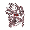
| ||||||||
| 4 | 
| ||||||||
| Unit cell |
|
-Components
| #1: Protein | Mass: 48900.305 Da / Num. of mol.: 4 Source method: isolated from a genetically manipulated source Source: (gene. exp.) Serratia sp. (strain FS14) (bacteria), (gene. exp.) Saccharomyces cerevisiae (brewer's yeast)Gene: malE, SIN3 / Production host: Escherichia coli (E. coli) / References: UniProt: A0A4P1LXE0, UniProt: P22579#2: Polysaccharide | 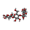  , Oligosaccharide / Class: Nutrient / Mass: 504.438 Da / Num. of mol.: 3 , Oligosaccharide / Class: Nutrient / Mass: 504.438 Da / Num. of mol.: 3Source method: isolated from a genetically manipulated source Details: oligosaccharide / References: beta-maltotriose #3: Polysaccharide | alpha-D-glucopyranose-(1-4)-alpha-D-glucopyranose / alpha-maltose |   , Oligosaccharide / Class: Nutrient / Mass: 342.297 Da / Num. of mol.: 1 , Oligosaccharide / Class: Nutrient / Mass: 342.297 Da / Num. of mol.: 1Source method: isolated from a genetically manipulated source Details: oligosaccharide / References: alpha-maltose #4: Water | ChemComp-HOH / | Water Mass: 18.015 Da / Num. of mol.: 702 / Source method: isolated from a natural source / Formula: H2O Mass: 18.015 Da / Num. of mol.: 702 / Source method: isolated from a natural source / Formula: H2OHas ligand of interest | N | |
|---|
-Experimental details
-Experiment
| Experiment | Method: X-RAY DIFFRACTION / Number of used crystals: 1 |
|---|
- Sample preparation
| Crystal | Density Matthews: 2.73 Å3/Da / Density % sol: 54.89 % |
|---|---|
| Crystal grow | Temperature: 293 K / Method: vapor diffusion, hanging drop / Details: 0.1M Bis-Tris, pH 6.0, 1.6M Ammonium sulfate |
-Data collection
| Diffraction | Mean temperature: 100 K / Serial crystal experiment: N | |||||||||||||||||||||
|---|---|---|---|---|---|---|---|---|---|---|---|---|---|---|---|---|---|---|---|---|---|---|
| Diffraction source | Source: SYNCHROTRON / Site: APS / Beamline: 24-ID-E / Wavelength: 0.9792 Å | |||||||||||||||||||||
| Detector | Type: DECTRIS PILATUS 6M-F / Detector: PIXEL / Date: Nov 12, 2019 | |||||||||||||||||||||
| Radiation | Protocol: SINGLE WAVELENGTH / Monochromatic (M) / Laue (L): M / Scattering type: x-ray | |||||||||||||||||||||
| Radiation wavelength | Wavelength: 0.9792 Å / Relative weight: 1 | |||||||||||||||||||||
| Reflection | Resolution: 2.2→75.841 Å / Num. obs: 105169 / % possible obs: 99.1 % / Redundancy: 3.5 % / Biso Wilson estimate: 39.692 Å2 / Rpim(I) all: 0.062 / Rrim(I) all: 0.117 / Net I/σ(I): 7.8 | |||||||||||||||||||||
| Reflection shell | Diffraction-ID: 1 / Redundancy: 3.4 %
|
-Phasing
| Phasing | Method: molecular replacement |
|---|
- Processing
| Software |
| ||||||||||||||||||||||||||||||||||||||||||||||||||||||||||||||||||||||||||||||||||||||||||||||||||||||||||||||||||||||||||||||||||||||||||||||||||||||||||||||||||||||||||||||||||||||||||
|---|---|---|---|---|---|---|---|---|---|---|---|---|---|---|---|---|---|---|---|---|---|---|---|---|---|---|---|---|---|---|---|---|---|---|---|---|---|---|---|---|---|---|---|---|---|---|---|---|---|---|---|---|---|---|---|---|---|---|---|---|---|---|---|---|---|---|---|---|---|---|---|---|---|---|---|---|---|---|---|---|---|---|---|---|---|---|---|---|---|---|---|---|---|---|---|---|---|---|---|---|---|---|---|---|---|---|---|---|---|---|---|---|---|---|---|---|---|---|---|---|---|---|---|---|---|---|---|---|---|---|---|---|---|---|---|---|---|---|---|---|---|---|---|---|---|---|---|---|---|---|---|---|---|---|---|---|---|---|---|---|---|---|---|---|---|---|---|---|---|---|---|---|---|---|---|---|---|---|---|---|---|---|---|---|---|---|---|
| Refinement | Method to determine structure: MOLECULAR REPLACEMENT Starting model: 4JBZ Resolution: 2.2→75.841 Å / SU ML: 0.32 / Cross valid method: THROUGHOUT / σ(F): 1.34 / Phase error: 27.46 / Stereochemistry target values: ML
| ||||||||||||||||||||||||||||||||||||||||||||||||||||||||||||||||||||||||||||||||||||||||||||||||||||||||||||||||||||||||||||||||||||||||||||||||||||||||||||||||||||||||||||||||||||||||||
| Solvent computation | Shrinkage radii: 0.9 Å / VDW probe radii: 1.11 Å / Solvent model: FLAT BULK SOLVENT MODEL | ||||||||||||||||||||||||||||||||||||||||||||||||||||||||||||||||||||||||||||||||||||||||||||||||||||||||||||||||||||||||||||||||||||||||||||||||||||||||||||||||||||||||||||||||||||||||||
| Displacement parameters | Biso max: 101.71 Å2 / Biso mean: 51.6779 Å2 / Biso min: 30.94 Å2 | ||||||||||||||||||||||||||||||||||||||||||||||||||||||||||||||||||||||||||||||||||||||||||||||||||||||||||||||||||||||||||||||||||||||||||||||||||||||||||||||||||||||||||||||||||||||||||
| Refinement step | Cycle: final / Resolution: 2.2→75.841 Å
| ||||||||||||||||||||||||||||||||||||||||||||||||||||||||||||||||||||||||||||||||||||||||||||||||||||||||||||||||||||||||||||||||||||||||||||||||||||||||||||||||||||||||||||||||||||||||||
| Refine LS restraints |
| ||||||||||||||||||||||||||||||||||||||||||||||||||||||||||||||||||||||||||||||||||||||||||||||||||||||||||||||||||||||||||||||||||||||||||||||||||||||||||||||||||||||||||||||||||||||||||
| LS refinement shell | Refine-ID: X-RAY DIFFRACTION / Rfactor Rfree error: 0
| ||||||||||||||||||||||||||||||||||||||||||||||||||||||||||||||||||||||||||||||||||||||||||||||||||||||||||||||||||||||||||||||||||||||||||||||||||||||||||||||||||||||||||||||||||||||||||
| Refinement TLS params. | Method: refined / Origin x: 39.6121 Å / Origin y: 100.595 Å / Origin z: 115.2588 Å
| ||||||||||||||||||||||||||||||||||||||||||||||||||||||||||||||||||||||||||||||||||||||||||||||||||||||||||||||||||||||||||||||||||||||||||||||||||||||||||||||||||||||||||||||||||||||||||
| Refinement TLS group |
|
