 Movie
Movie Controller


+ Open data

- Basic information
| Entry | Database: PDB / ID: 6x4h | ||||||
|---|---|---|---|---|---|---|---|
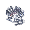
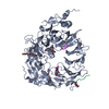
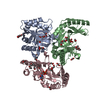
| Title | Sortilin-Progranulin Interaction With Compound 24 | ||||||
 Components Components | Sortilin | ||||||
 Keywords Keywords | UNKNOWN FUNCTION /  Protein Protein Interaction Protein Protein Interaction | ||||||
| Function / homology |  Function and homology information Function and homology informationneurotensin receptor activity, non-G protein-coupled / negative regulation of lipoprotein lipase activity / Golgi to lysosome transport / myotube differentiation / cerebellar climbing fiber to Purkinje cell synapse / plasma membrane to endosome transport / retromer complex binding / maintenance of synapse structure / Golgi to endosome transport / nerve growth factor receptor activity ...neurotensin receptor activity, non-G protein-coupled / negative regulation of lipoprotein lipase activity / Golgi to lysosome transport / myotube differentiation / cerebellar climbing fiber to Purkinje cell synapse / plasma membrane to endosome transport / retromer complex binding / maintenance of synapse structure / Golgi to endosome transport / nerve growth factor receptor activity / vesicle organization / endosome transport via multivesicular body sorting pathway / nerve growth factor binding / protein targeting to lysosome / trans-Golgi network transport vesicle / clathrin-coated vesicle / Golgi cisterna membrane / Golgi Associated Vesicle Biogenesis / negative regulation of fat cell differentiation / endosome to lysosome transport / glucose import / neurotrophin TRK receptor signaling pathway / extrinsic apoptotic signaling pathway via death domain receptors / neuropeptide signaling pathway / clathrin-coated pit / ossification / response to insulin / endocytosis / cytoplasmic vesicle / regulation of gene expression / nuclear membrane / lysosome / early endosome / endosome membrane / G protein-coupled receptor signaling pathway / lysosomal membrane / endoplasmic reticulum membrane / perinuclear region of cytoplasm / Golgi apparatus / enzyme binding / cell surface / membrane / plasma membrane / cytosolSimilarity search - Function | ||||||
| Biological species |  Homo sapiens (human) Homo sapiens (human) | ||||||
| Method | X-RAY DIFFRACTION / SYNCHROTRON / MOLECULAR REPLACEMENT / Resolution: 2.9 Å | ||||||
 Authors Authors | Parthasarathy, G. / Soisson, S.M. / Klein, D. | ||||||
 Citation Citation | Journal: Bioorg.Med.Chem.Lett. / Year: 2020 Title: Identification of potent inhibitors of the sortilin-progranulin interaction. Authors: Stachel, S.J. / Ginnetti, A.T. / Johnson, S.A. / Cramer, P. / Wang, Y. / Bukhtiyarova, M. / Krosky, D. / Stump, C. / Hurzy, D.M. / Schlegel, K.A. / Cooke, A.J. / Allen, S. / O'Donnell, G. / ...Authors: Stachel, S.J. / Ginnetti, A.T. / Johnson, S.A. / Cramer, P. / Wang, Y. / Bukhtiyarova, M. / Krosky, D. / Stump, C. / Hurzy, D.M. / Schlegel, K.A. / Cooke, A.J. / Allen, S. / O'Donnell, G. / Ziebell, M. / Parthasarathy, G. / Getty, K.L. / Ho, T. / Ou, Y. / Jovanovska, A. / Carroll, S.S. / Pausch, M. / Lumb, K. / Mosser, S.D. / Voleti, B. / Klein, D.J. / Soisson, S.M. / Zerbinatti, C. / Coleman, P.J. | ||||||
| History |
|
- Structure visualization
| Structure viewer | Molecule: MolmilJmol/JSmol |
|---|
- Downloads & links
-Download
| PDBx/mmCIF format | 6x4h.cif.gz | 146.3 KB | Display | PDBx/mmCIF format |
|---|---|---|---|---|
| PDB format | pdb6x4h.ent.gz | 110.8 KB | Display | PDB format |
| PDBx/mmJSON format | 6x4h.json.gz | Tree view | PDBx/mmJSON format | |
| Others |  Other downloads Other downloads |
-Validation report
| Arichive directory | https://data.pdbj.org/pub/pdb/validation_reports/x4/6x4hftp://data.pdbj.org/pub/pdb/validation_reports/x4/6x4h | HTTPS FTP |
|---|
-Related structure data
| Related structure data |  6x3lC  6x48C  3f6kS S: Starting model for refinement C: citing same article ( |
|---|---|
| Similar structure data |


-Links
 PDBj
PDBj
- Assembly
| Deposited unit | 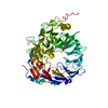
| ||||||||
|---|---|---|---|---|---|---|---|---|---|
| 1 |
| ||||||||
| Unit cell |
|
-Components
| #1: Protein | Mass: 74255.133 Da / Num. of mol.: 1 Source method: isolated from a genetically manipulated source Source: (gene. exp.) Homo sapiens (human) / Gene: SORT1 / Production host:   Cricetulus griseus (Chinese hamster) / References: UniProt: Q99523 Cricetulus griseus (Chinese hamster) / References: UniProt: Q99523 | ||||||||
|---|---|---|---|---|---|---|---|---|---|
| #2: Polysaccharide | / Mass: 424.401 Da / Num. of mol.: 2 Source method: isolated from a genetically manipulated source #3: Chemical | ChemComp-UOY / |   Mass: 342.389 Da / Num. of mol.: 1 / Source method: obtained synthetically / Formula: C19H22N2O4 / Feature type: SUBJECT OF INVESTIGATION Mass: 342.389 Da / Num. of mol.: 1 / Source method: obtained synthetically / Formula: C19H22N2O4 / Feature type: SUBJECT OF INVESTIGATION#4: Chemical | ChemComp-GOL / | Glycerol  Mass: 92.094 Da / Num. of mol.: 1 / Source method: obtained synthetically / Formula: C3H8O3 / Feature type: SUBJECT OF INVESTIGATION Mass: 92.094 Da / Num. of mol.: 1 / Source method: obtained synthetically / Formula: C3H8O3 / Feature type: SUBJECT OF INVESTIGATION#5: Water | ChemComp-HOH / | Water Mass: 18.015 Da / Num. of mol.: 10 / Source method: isolated from a natural source / Formula: H2O Mass: 18.015 Da / Num. of mol.: 10 / Source method: isolated from a natural source / Formula: H2OHas ligand of interest | Y | |
-Experimental details
-Experiment
| Experiment | Method: X-RAY DIFFRACTION / Number of used crystals: 1 |
|---|
- Sample preparation
| Crystal | Density Matthews: 3.9 Å3/Da / Density % sol: 68.49 % |
|---|---|
| Crystal grow | Temperature: 293 K / Method: vapor diffusion, hanging drop Details: 20-25% PEG3350, 0.4M Na Malonate, pH8, 0.1M Tris, 5% Glycerol |
-Data collection
| Diffraction | Mean temperature: 100 K / Serial crystal experiment: N |
|---|---|
| Diffraction source | Source: SYNCHROTRON / Site: APS  / Beamline: 17-ID / Wavelength: 1 Å / Beamline: 17-ID / Wavelength: 1 Å |
| Detector | Type: DECTRIS PILATUS3 6M / Detector: PIXEL / Date: Jun 20, 2014 |
| Radiation | Protocol: SINGLE WAVELENGTH / Monochromatic (M) / Laue (L): M / Scattering type: x-ray |
| Radiation wavelength | Wavelength: 1 Å / Relative weight: 1 |
| Reflection | Resolution: 2.9→89 Å / Num. obs: 24571 / % possible obs: 97.9 % / Redundancy: 3.3 % / Rmerge(I) obs: 0.054 / Net I/σ(I): 12.9 |
| Reflection shell | Resolution: 2.904→2.913 Å / Rmerge(I) obs: 0.522 / Num. unique obs: 231 |
- Processing
| Software |
| ||||||||||||||||||||||||||||||||||||||||||||||||||||||||||||||||||||||||||||||||||||||||||||||||||||||||||||
|---|---|---|---|---|---|---|---|---|---|---|---|---|---|---|---|---|---|---|---|---|---|---|---|---|---|---|---|---|---|---|---|---|---|---|---|---|---|---|---|---|---|---|---|---|---|---|---|---|---|---|---|---|---|---|---|---|---|---|---|---|---|---|---|---|---|---|---|---|---|---|---|---|---|---|---|---|---|---|---|---|---|---|---|---|---|---|---|---|---|---|---|---|---|---|---|---|---|---|---|---|---|---|---|---|---|---|---|---|---|
| Refinement | Method to determine structure: MOLECULAR REPLACEMENT Starting model: 3F6K Resolution: 2.9→32.29 Å / Cor.coef. Fo:Fc: 0.9223 / Cor.coef. Fo:Fc free: 0.8906 / SU R Cruickshank DPI: 0.708 / Cross valid method: THROUGHOUT / σ(F): 0 / SU R Blow DPI: 0.617 / SU Rfree Blow DPI: 0.328 / SU Rfree Cruickshank DPI: 0.34
| ||||||||||||||||||||||||||||||||||||||||||||||||||||||||||||||||||||||||||||||||||||||||||||||||||||||||||||
| Displacement parameters | Biso max: 186.41 Å2 / Biso mean: 92.59 Å2 / Biso min: 40.56 Å2
| ||||||||||||||||||||||||||||||||||||||||||||||||||||||||||||||||||||||||||||||||||||||||||||||||||||||||||||
| Refine analyze | Luzzati coordinate error obs: 0.417 Å | ||||||||||||||||||||||||||||||||||||||||||||||||||||||||||||||||||||||||||||||||||||||||||||||||||||||||||||
| Refinement step | Cycle: final / Resolution: 2.9→32.29 Å
| ||||||||||||||||||||||||||||||||||||||||||||||||||||||||||||||||||||||||||||||||||||||||||||||||||||||||||||
| Refine LS restraints |
| ||||||||||||||||||||||||||||||||||||||||||||||||||||||||||||||||||||||||||||||||||||||||||||||||||||||||||||
| LS refinement shell | Resolution: 2.9→3.03 Å / Total num. of bins used: 12
|
