National Institutes of Health/National Institute of Neurological Disorders and Stroke (NIH/NINDS)
R01NS108151
United States
National Institutes of Health/National Institute of General Medical Sciences (NIH/NIGMS)
R01GM121994
United States
National Institutes of Health/National Institute of Diabetes and Digestive and Kidney Disease (NIH/NIDDK)
R01DK099023
United States
National Institutes of Health/National Institute of General Medical Sciences (NIH/NIGMS)
United States
Citation
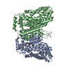
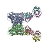
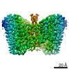
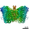
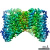
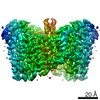
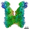
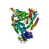
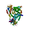
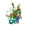
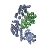
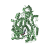
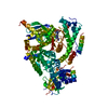
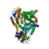
Journal: Elife / Year: 2020 Title: Structural basis for the reaction cycle of DASS dicarboxylate transporters. Authors: David B Sauer / Noah Trebesch / Jennifer J Marden / Nicolette Cocco / Jinmei Song / Akiko Koide / Shohei Koide / Emad Tajkhorshid / Da-Neng Wang / Abstract: Citrate, α-ketoglutarate and succinate are TCA cycle intermediates that also play essential roles in metabolic signaling and cellular regulation. These di- and tricarboxylates are imported into the ...Citrate, α-ketoglutarate and succinate are TCA cycle intermediates that also play essential roles in metabolic signaling and cellular regulation. These di- and tricarboxylates are imported into the cell by the divalent anion sodium symporter (DASS) family of plasma membrane transporters, which contains both cotransporters and exchangers. While DASS proteins transport substrates via an elevator mechanism, to date structures are only available for a single DASS cotransporter protein in a substrate-bound, inward-facing state. We report multiple cryo-EM and X-ray structures in four different states, including three hitherto unseen states, along with molecular dynamics simulations, of both a cotransporter and an exchanger. Comparison of these outward- and inward-facing structures reveal how the transport domain translates and rotates within the framework of the scaffold domain through the transport cycle. Additionally, we propose that DASS transporters ensure substrate coupling by a charge-compensation mechanism, and by structural changes upon substrate release.
Component-ID: 1 / Ens-ID: 1 / Beg auth comp-ID: ALA / Beg label comp-ID: ALA / End auth comp-ID: ILE / End label comp-ID: ILE / Auth seq-ID: 0 - 487 / Label seq-ID: 3 - 490
Dom-ID
Selection details
Auth asym-ID
Label asym-ID
1
(chain 'A' and (resid0through265or (resid266...
A
A
2
chain 'B'
B
B
-
Components
#1: Protein
DASSfamilysodium-coupledanionsymporter
Mass: 53539.527 Da / Num. of mol.: 2 Source method: isolated from a genetically manipulated source Source: (gene. exp.) Lactobacillus acidophilus (bacteria) / Gene: D7H66_01865 / Production host: Escherichia coli (E. coli) / References: UniProt: A0A3A9Y7Q2, UniProt: Q5FKK5*PLUS
-
Experimental details
-
Experiment
Experiment
Method: X-RAY DIFFRACTION / Number of used crystals: 1
-
Sample preparation
Crystal
Density Matthews: 3 Å3/Da / Density % sol: 58.97 %
Crystal grow
Temperature: 291 K / Method: vapor diffusion, hanging drop / pH: 6.8 / Details: 30% Jeffamine ED-2001, 100 mM HEPES pH 6.8
-
Data collection
Diffraction
Mean temperature: 100 K / Serial crystal experiment: N
Diffraction source
Source: SYNCHROTRON / Site: ALS / Beamline: 5.0.2 / Wavelength: 1 Å
In the structure databanks used in Yorodumi, some data are registered as the other names, "COVID-19 virus" and "2019-nCoV". Here are the details of the virus and the list of structure data.
Jan 31, 2019. EMDB accession codes are about to change! (news from PDBe EMDB page)
EMDB accession codes are about to change! (news from PDBe EMDB page)
The allocation of 4 digits for EMDB accession codes will soon come to an end. Whilst these codes will remain in use, new EMDB accession codes will include an additional digit and will expand incrementally as the available range of codes is exhausted. The current 4-digit format prefixed with “EMD-” (i.e. EMD-XXXX) will advance to a 5-digit format (i.e. EMD-XXXXX), and so on. It is currently estimated that the 4-digit codes will be depleted around Spring 2019, at which point the 5-digit format will come into force.
The EM Navigator/Yorodumi systems omit the EMD- prefix.
Related info.:Q: What is EMD? / ID/Accession-code notation in Yorodumi/EM Navigator
Yorodumi is a browser for structure data from EMDB, PDB, SASBDB, etc.
This page is also the successor to EM Navigator detail page, and also detail information page/front-end page for Omokage search.
The word "yorodu" (or yorozu) is an old Japanese word meaning "ten thousand". "mi" (miru) is to see.
Related info.:EMDB / PDB / SASBDB / Comparison of 3 databanks / Yorodumi Search / Aug 31, 2016. New EM Navigator & Yorodumi / Yorodumi Papers / Jmol/JSmol / Function and homology information / Changes in new EM Navigator and Yorodumi
 Movie
Movie Controller

 Yorodumi
 Open data
 Basic information
 Components
 Keywords
 TRANSPORT PROTEIN /
  Function and homology information
 Authors
 United States, 4items
  Citation
 Structure visualization
 Downloads & links
 Other downloads
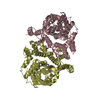


 PDBj
PDBj Assembly
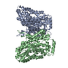
 Sample preparation
 Processing