 Movie
Movie Controller

[English] 日本語
 Yorodumi
- PDB-6wsj: Crystal Structure of Danio rerio histone deacetylase 6 catalytic ... -
+ Open data

- Basic information
| Entry | Database: PDB / ID: 6wsj | ||||||
|---|---|---|---|---|---|---|---|
| Title | Crystal Structure of Danio rerio histone deacetylase 6 catalytic domain 2 complexed with cyclopeptide des4.3.1 | ||||||
 Components Components |
| ||||||
 Keywords Keywords | hydrolase/hydrolase inhibitor /  Hydrolase / histone deacetylase / inhibitor / metallohydrolase / hydrolase-hydrolase inhibitor complex Hydrolase / histone deacetylase / inhibitor / metallohydrolase / hydrolase-hydrolase inhibitor complex | ||||||
| Function / homology |  Function and homology information Function and homology informationtubulin deacetylase activity / swimming behavior / definitive hemopoiesis / protein lysine deacetylase activity / histone deacetylase activity / regulation of tubulin deacetylation / potassium ion binding / histone deacetylase complex / hematopoietic progenitor cell differentiation / angiogenesis ...tubulin deacetylase activity / swimming behavior / definitive hemopoiesis / protein lysine deacetylase activity / histone deacetylase activity / regulation of tubulin deacetylation / potassium ion binding / histone deacetylase complex / hematopoietic progenitor cell differentiation / angiogenesis / negative regulation of transcription by RNA polymerase II / zinc ion bindingSimilarity search - Function | ||||||
| Biological species |  Danio rerio (zebrafish) Danio rerio (zebrafish)synthetic construct (others) | ||||||
| Method | X-RAY DIFFRACTION / SYNCHROTRON / MOLECULAR REPLACEMENT / Resolution: 1.7 Å | ||||||
 Authors Authors | Watson, P.R. / Christianson, D.W. | ||||||
| Funding support |  United States, 1items United States, 1items
| ||||||
 Citation Citation | Journal: Nat Commun / Year: 2021 Title: Anchor extension: a structure-guided approach to design cyclic peptides targeting enzyme active sites. Authors: Hosseinzadeh, P. / Watson, P.R. / Craven, T.W. / Li, X. / Rettie, S. / Pardo-Avila, F. / Bera, A.K. / Mulligan, V.K. / Lu, P. / Ford, A.S. / Weitzner, B.D. / Stewart, L.J. / Moyer, A.P. / Di ...Authors: Hosseinzadeh, P. / Watson, P.R. / Craven, T.W. / Li, X. / Rettie, S. / Pardo-Avila, F. / Bera, A.K. / Mulligan, V.K. / Lu, P. / Ford, A.S. / Weitzner, B.D. / Stewart, L.J. / Moyer, A.P. / Di Piazza, M. / Whalen, J.G. / Greisen, P.J. / Christianson, D.W. / Baker, D. | ||||||
| History |
|
- Structure visualization
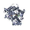
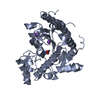
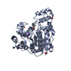
| Structure viewer | Molecule: MolmilJmol/JSmol |
|---|
- Downloads & links
-Download
| PDBx/mmCIF format | 6wsj.cif.gz | 113.5 KB | Display | PDBx/mmCIF format |
|---|---|---|---|---|
| PDB format | pdb6wsj.ent.gz | 67.7 KB | Display | PDB format |
| PDBx/mmJSON format | 6wsj.json.gz | Tree view | PDBx/mmJSON format | |
| Others |  Other downloads Other downloads |
-Validation report
| Arichive directory | https://data.pdbj.org/pub/pdb/validation_reports/ws/6wsjftp://data.pdbj.org/pub/pdb/validation_reports/ws/6wsj | HTTPS FTP |
|---|
-Related structure data
| Related structure data |  6whnC  6whoC  6whqC  6whzC  6wi3C 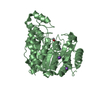 5eemS S: Starting model for refinement C: citing same article ( |
|---|---|
| Similar structure data |


-Links
 PDBj
PDBj- Assembly
| Deposited unit | 
| ||||||||||||
|---|---|---|---|---|---|---|---|---|---|---|---|---|---|
| 1 |
| ||||||||||||
| Unit cell |
|
-Components
-Protein / Protein/peptide , 2 types, 2 molecules AI
| #1: Protein | / Histone Deacetylase 6 Mass: 40285.484 Da / Num. of mol.: 1 Source method: isolated from a genetically manipulated source Source: (gene. exp.) Danio rerio (zebrafish) / Gene: hdac6 / Production host:  Escherichia coli (E. coli) / References: UniProt: A7YT55 Escherichia coli (E. coli) / References: UniProt: A7YT55 |
|---|---|
| #2: Protein/peptide |   Type: Cyclic peptide / Class: Enzyme inhibitor / Mass: 997.213 Da / Num. of mol.: 1 / Source method: obtained synthetically / Source: (synth.) synthetic construct (others) / References: des4.3.1 Type: Cyclic peptide / Class: Enzyme inhibitor / Mass: 997.213 Da / Num. of mol.: 1 / Source method: obtained synthetically / Source: (synth.) synthetic construct (others) / References: des4.3.1 |
-Non-polymers , 4 types, 244 molecules 


| #3: Chemical | ChemComp-ZN /  Mass: 65.409 Da / Num. of mol.: 1 / Source method: obtained synthetically / Formula: Zn Mass: 65.409 Da / Num. of mol.: 1 / Source method: obtained synthetically / Formula: Zn | ||||
|---|---|---|---|---|---|
| #4: Chemical |  Mass: 39.098 Da / Num. of mol.: 2 / Source method: obtained synthetically / Formula: K Mass: 39.098 Da / Num. of mol.: 2 / Source method: obtained synthetically / Formula: K#5: Chemical | Ethylene glycol Mass: 62.068 Da / Num. of mol.: 2 / Source method: obtained synthetically / Formula: C2H6O2 Mass: 62.068 Da / Num. of mol.: 2 / Source method: obtained synthetically / Formula: C2H6O2#6: Water | ChemComp-HOH / | WaterMass: 18.015 Da / Num. of mol.: 239 / Source method: isolated from a natural source / Formula: H2O |
-Details
| Has ligand of interest | Y |
|---|
-Experimental details
-Experiment
| Experiment | Method: X-RAY DIFFRACTION / Number of used crystals: 1 |
|---|
- Sample preparation
| Crystal | Density Matthews: 2.53 Å3/Da / Density % sol: 51.48 % |
|---|---|
| Crystal grow | Temperature: 277 K / Method: vapor diffusion, sitting drop Details: 10 mg/mL HDAC6 protein 0.2 M ammonium chloride and 20% polyethylene glycol (PEG) 3350 (w/v) |
-Data collection
| Diffraction | Mean temperature: 100 K / Serial crystal experiment: N |
|---|---|
| Diffraction source | Source: SYNCHROTRON / Site: NSLS-II / Beamline: 17-ID-1 / Wavelength: 0.98 Å |
| Detector | Type: DECTRIS EIGER X 9M / Detector: PIXEL / Date: Feb 12, 2020 |
| Radiation | Protocol: SINGLE WAVELENGTH / Monochromatic (M) / Laue (L): M / Scattering type: x-ray |
| Radiation wavelength | Wavelength: 0.98 Å / Relative weight: 1 |
| Reflection | Resolution: 1.7→62.76 Å / Num. obs: 45672 / % possible obs: 100 % / Redundancy: 6.4 % / Biso Wilson estimate: 12.48 Å2 / CC1/2: 0.994 / Rmerge(I) obs: 0.126 / Rpim(I) all: 0.079 / Net I/σ(I): 9.8 |
| Reflection shell | Resolution: 1.7→1.76 Å / Rmerge(I) obs: 0.619 / Mean I/σ(I) obs: 3.8 / Num. unique obs: 45672 / CC1/2: 0.845 / Rpim(I) all: 0.413 |
- Processing
| Software |
| |||||||||||||||||||||||||||||||||||||||||||||||||||||||||||||||||||||||||||||||||||||||||||||||||||||||||||||||||||||||
|---|---|---|---|---|---|---|---|---|---|---|---|---|---|---|---|---|---|---|---|---|---|---|---|---|---|---|---|---|---|---|---|---|---|---|---|---|---|---|---|---|---|---|---|---|---|---|---|---|---|---|---|---|---|---|---|---|---|---|---|---|---|---|---|---|---|---|---|---|---|---|---|---|---|---|---|---|---|---|---|---|---|---|---|---|---|---|---|---|---|---|---|---|---|---|---|---|---|---|---|---|---|---|---|---|---|---|---|---|---|---|---|---|---|---|---|---|---|---|---|---|
| Refinement | Method to determine structure: MOLECULAR REPLACEMENT Starting model: PDB Entry 5EEM Resolution: 1.7→62.76 Å / SU ML: 0.1623 / Cross valid method: FREE R-VALUE / σ(F): 1.34 / Phase error: 15.3393 / Stereochemistry target values: GeoStd + Monomer Library
| |||||||||||||||||||||||||||||||||||||||||||||||||||||||||||||||||||||||||||||||||||||||||||||||||||||||||||||||||||||||
| Solvent computation | Shrinkage radii: 0.9 Å / VDW probe radii: 1.11 Å / Solvent model: FLAT BULK SOLVENT MODEL | |||||||||||||||||||||||||||||||||||||||||||||||||||||||||||||||||||||||||||||||||||||||||||||||||||||||||||||||||||||||
| Displacement parameters | Biso mean: 12.88 Å2 | |||||||||||||||||||||||||||||||||||||||||||||||||||||||||||||||||||||||||||||||||||||||||||||||||||||||||||||||||||||||
| Refinement step | Cycle: LAST / Resolution: 1.7→62.76 Å
| |||||||||||||||||||||||||||||||||||||||||||||||||||||||||||||||||||||||||||||||||||||||||||||||||||||||||||||||||||||||
| Refine LS restraints |
| |||||||||||||||||||||||||||||||||||||||||||||||||||||||||||||||||||||||||||||||||||||||||||||||||||||||||||||||||||||||
| LS refinement shell |
|
