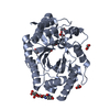
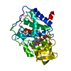
 Movie
Movie Controller


+ Open data

- Basic information
| Entry | Database: PDB / ID: 6wqw | ||||||||||||
|---|---|---|---|---|---|---|---|---|---|---|---|---|---|
| Title | Thermobacillus composti GH10 xylanase | ||||||||||||
 Components Components | Beta-xylanase Xylanase Xylanase | ||||||||||||
 Keywords Keywords | HYDROLASE / GH 10 / Glycoside Hydrolase / Thermobacillus composti | ||||||||||||
| Function / homology |  Function and homology informationendo-1,4-beta-xylanase activity / endo-1,4-beta-xylanase / xylan catabolic process Function and homology informationendo-1,4-beta-xylanase activity / endo-1,4-beta-xylanase / xylan catabolic processSimilarity search - Function | ||||||||||||
| Biological species |  Thermobacillus composti (bacteria) Thermobacillus composti (bacteria) | ||||||||||||
| Method | X-RAY DIFFRACTION / SYNCHROTRON / MOLECULAR REPLACEMENT / molecular replacement / Resolution: 2.102 Å | ||||||||||||
 Authors Authors | Briganti, L. / Polikarpov, I. | ||||||||||||
| Funding support |  Brazil, 3items Brazil, 3items
| ||||||||||||
 Citation Citation | Journal: Carbohydr Polym / Year: 2020 Title: Transformation of xylan into value-added biocommodities using Thermobacillus composti GH10 xylanase. Authors: Sepulchro, A.G.V. / Pellegrini, V.O.A. / Briganti, L. / de Araujo, E.A. / de Araujo, S.S. / Polikarpov, I. | ||||||||||||
| History |
|
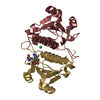
- Structure visualization
| Structure viewer | Molecule: MolmilJmol/JSmol |
|---|
- Downloads & links
-Download
| PDBx/mmCIF format | 6wqw.cif.gz | 142.1 KB | Display | PDBx/mmCIF format |
|---|---|---|---|---|
| PDB format | pdb6wqw.ent.gz | 110.5 KB | Display | PDB format |
| PDBx/mmJSON format | 6wqw.json.gz | Tree view | PDBx/mmJSON format | |
| Others |  Other downloads Other downloads |
-Validation report
| Arichive directory | https://data.pdbj.org/pub/pdb/validation_reports/wq/6wqwftp://data.pdbj.org/pub/pdb/validation_reports/wq/6wqw | HTTPS FTP |
|---|
-Related structure data
| Related structure data | 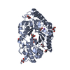 1n82S S: Starting model for refinement |
|---|---|
| Similar structure data |


-Links
 PDBj
PDBj
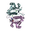
- Assembly
| Deposited unit | 
| ||||||||
|---|---|---|---|---|---|---|---|---|---|
| 1 |
| ||||||||
| Unit cell |
|
-Components
| #1: Protein | Xylanase Mass: 38594.414 Da / Num. of mol.: 1 Source method: isolated from a genetically manipulated source Source: (gene. exp.) Thermobacillus composti (bacteria) / Production host: Escherichia coli (E. coli) / References: UniProt: L0EGW1, endo-1,4-beta-xylanase |
|---|---|
| #2: Sugar | ChemComp-XYP / Xylose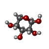  Type: D-saccharide, beta linking / Mass: 150.130 Da / Num. of mol.: 1 Type: D-saccharide, beta linking / Mass: 150.130 Da / Num. of mol.: 1Source method: isolated from a genetically manipulated source Formula: C5H10O5 / Feature type: SUBJECT OF INVESTIGATION |
| #3: Water | ChemComp-HOH / Water Mass: 18.015 Da / Num. of mol.: 106 / Source method: isolated from a natural source / Formula: H2O Mass: 18.015 Da / Num. of mol.: 106 / Source method: isolated from a natural source / Formula: H2O |
| Has ligand of interest | Y |
-Experimental details
-Experiment
| Experiment | Method: X-RAY DIFFRACTION / Number of used crystals: 1 |
|---|
- Sample preparation
| Crystal | Density Matthews: 1.98 Å3/Da / Density % sol: 37.74 % |
|---|---|
| Crystal grow | Temperature: 293 K / Method: vapor diffusion, sitting drop / pH: 7.5 Details: 0.1 M HEPES, 70% (v/v) 2-Methyl-2,4-pentanediol (MPD) |
-Data collection
| Diffraction | Mean temperature: 100 K / Serial crystal experiment: N |
|---|---|
| Diffraction source | Source: SYNCHROTRON / Site: LNLS / Beamline: W01B-MX2 / Wavelength: 1.4586 Å |
| Detector | Type: DECTRIS PILATUS 2M / Detector: PIXEL / Date: May 10, 2019 |
| Radiation | Protocol: SINGLE WAVELENGTH / Monochromatic (M) / Laue (L): M / Scattering type: x-ray |
| Radiation wavelength | Wavelength: 1.4586 Å / Relative weight: 1 |
| Reflection | Resolution: 2.102→33.82 Å / Num. obs: 18595 / % possible obs: 98.4 % / Redundancy: 22.7 % / CC1/2: 1 / Net I/σ(I): 18.26 |
| Reflection shell | Resolution: 2.102→2.18 Å / Redundancy: 19.4 % / Mean I/σ(I) obs: 2.23 / Num. unique obs: 1809 / CC1/2: 0.868 / % possible all: 99.78 |
-Phasing
| Phasing | Method: molecular replacement | |||||||||
|---|---|---|---|---|---|---|---|---|---|---|
| Phasing MR | Model details: Phaser MODE: MR_AUTO
|
- Processing
| Software |
| ||||||||||||||||||||||||||||||||||||||||||||||||
|---|---|---|---|---|---|---|---|---|---|---|---|---|---|---|---|---|---|---|---|---|---|---|---|---|---|---|---|---|---|---|---|---|---|---|---|---|---|---|---|---|---|---|---|---|---|---|---|---|---|
| Refinement | Method to determine structure: MOLECULAR REPLACEMENT Starting model: 1N82 Resolution: 2.102→33.819 Å / SU ML: 0.24 / Cross valid method: THROUGHOUT / σ(F): 1.33 / Phase error: 29.13 / Stereochemistry target values: ML
| ||||||||||||||||||||||||||||||||||||||||||||||||
| Solvent computation | Shrinkage radii: 0.9 Å / VDW probe radii: 1.11 Å / Solvent model: FLAT BULK SOLVENT MODEL | ||||||||||||||||||||||||||||||||||||||||||||||||
| Displacement parameters | Biso max: 109.23 Å2 / Biso mean: 48.2512 Å2 / Biso min: 16.26 Å2 | ||||||||||||||||||||||||||||||||||||||||||||||||
| Refinement step | Cycle: final / Resolution: 2.102→33.819 Å
| ||||||||||||||||||||||||||||||||||||||||||||||||
| LS refinement shell | Refine-ID: X-RAY DIFFRACTION / Rfactor Rfree error: 0
| ||||||||||||||||||||||||||||||||||||||||||||||||
| Refinement TLS params. | Method: refined / Origin x: 54.044 Å / Origin y: 25.2786 Å / Origin z: 47.0567 Å
| ||||||||||||||||||||||||||||||||||||||||||||||||
| Refinement TLS group | Selection details: (chain A and resseq 4:330) |
