National Institutes of Health/National Institute on Aging (NIH/NIA)
AG054022
米国
引用
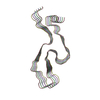
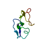
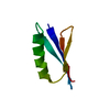
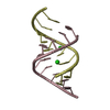
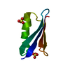
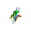
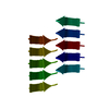
ジャーナル: Nat Commun / 年: 2020 タイトル: CryoEM structure of the low-complexity domain of hnRNPA2 and its conversion to pathogenic amyloid. 著者: Jiahui Lu / Qin Cao / Michael P Hughes / Michael R Sawaya / David R Boyer / Duilio Cascio / David S Eisenberg / 要旨: hnRNPA2 is a human ribonucleoprotein (RNP) involved in RNA metabolism. It forms fibrils both under cellular stress and in mutated form in neurodegenerative conditions. Previous work established that ...hnRNPA2 is a human ribonucleoprotein (RNP) involved in RNA metabolism. It forms fibrils both under cellular stress and in mutated form in neurodegenerative conditions. Previous work established that the C-terminal low-complexity domain (LCD) of hnRNPA2 fibrillizes under stress, and missense mutations in this domain are found in the disease multisystem proteinopathy (MSP). However, little is known at the atomic level about the hnRNPA2 LCD structure that is involved in those processes and how disease mutations cause structural change. Here we present the cryo-electron microscopy (cryoEM) structure of the hnRNPA2 LCD fibril core and demonstrate its capability to form a reversible hydrogel in vitro containing amyloid-like fibrils. Whereas these fibrils, like pathogenic amyloid, are formed from protein chains stacked into β-sheets by backbone hydrogen bonds, they display distinct structural differences: the chains are kinked, enabling non-covalent cross-linking of fibrils and disfavoring formation of pathogenic steric zippers. Both reversibility and energetic calculations suggest these fibrils are less stable than pathogenic amyloid. Moreover, the crystal structure of the disease-mutation-containing segment (D290V) of hnRNPA2 suggests that the replacement fundamentally alters the fibril structure to a more stable energetic state. These findings illuminate how molecular interactions promote protein fibril networks and how mutation can transform fibril structure from functional to a pathogenic form.
構造決定の手法: AB INITIO PHASING / 解像度: 1.1→20.64 Å / Cor.coef. Fo:Fc: 0.992 / Cor.coef. Fo:Fc free: 0.99 / SU B: 0.676 / SU ML: 0.014 / SU R Cruickshank DPI: 0.0245 / 交差検証法: THROUGHOUT / σ(F): 0 / ESU R: 0.025 / ESU R Free: 0.026 詳細: HYDROGENS HAVE BEEN USED IF PRESENT IN THE INPUT U VALUES : REFINED INDIVIDUALLY
 ムービー
ムービー コントローラー


 データを開く
 基本情報
基本情報 要素
要素 キーワード
キーワード 機能・相同性情報
 カハール体 / mRNA export from nucleus / pre-mRNA intronic binding / Gene and protein expression by JAK-STAT signaling after Interleukin-12 stimulation / catalytic step 2 spliceosome / mRNA Splicing - Major Pathway / molecular condensate scaffold activity / mRNA 3'-UTR binding /
 データ登録者
 米国, 2件
  引用
引用 構造の表示
構造の表示 ダウンロードとリンク
 その他のダウンロード


 PDBj
PDBj


 集合体
集合体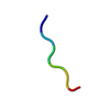
 分子量: 18.015 Da / 分子数: 3 / 由来タイプ: 天然 / 式: H2O
 試料調製
試料調製 解析
解析