 Movie
Movie Controller

[English] 日本語
 Yorodumi
- PDB-6wpq: GNYNVF from hnRNPA2-low complexity domain segment, residues 286-2... -
+ Open data

- Basic information
| Entry | Database: PDB / ID: 6wpq | |||||||||
|---|---|---|---|---|---|---|---|---|---|---|
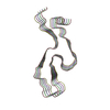
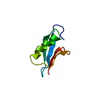
| Title | GNYNVF from hnRNPA2-low complexity domain segment, residues 286-291, D290V variant | |||||||||
 Components Components | Heterogeneous nuclear ribonucleoprotein A2 | |||||||||
 Keywords Keywords | PROTEIN FIBRIL / amyloidogenic segment / amyloid-like fibril / disease related / multisystem proteinopathy (MSP) | |||||||||
| Function / homology |  Function and homology information Function and homology informationpositive regulation of telomerase RNA reverse transcriptase activity / miRNA transport / positive regulation of telomere maintenance via telomere lengthening / RNA transport / G-quadruplex DNA unwinding / primary miRNA processing / single-stranded telomeric DNA binding / N6-methyladenosine-containing RNA reader activity / G-rich strand telomeric DNA binding / miRNA binding ...positive regulation of telomerase RNA reverse transcriptase activity / miRNA transport / positive regulation of telomere maintenance via telomere lengthening / RNA transport / G-quadruplex DNA unwinding / primary miRNA processing / single-stranded telomeric DNA binding / N6-methyladenosine-containing RNA reader activity / G-rich strand telomeric DNA binding / miRNA binding / negative regulation of mRNA splicing, via spliceosome / Processing of Capped Intron-Containing Pre-mRNA / mRNA transport /  Cajal body / mRNA export from nucleus / pre-mRNA intronic binding / Gene and protein expression by JAK-STAT signaling after Interleukin-12 stimulation / catalytic step 2 spliceosome / molecular condensate scaffold activity / mRNA Splicing - Major Pathway / mRNA 3'-UTR binding / spliceosomal complex / mRNA processing / mRNA splicing, via spliceosome / nuclear matrix / chromosome, telomeric region / ribonucleoprotein complex / negative regulation of transcription by RNA polymerase II / RNA binding / extracellular exosome / nucleoplasm / membrane / identical protein binding / nucleus / cytoplasm Cajal body / mRNA export from nucleus / pre-mRNA intronic binding / Gene and protein expression by JAK-STAT signaling after Interleukin-12 stimulation / catalytic step 2 spliceosome / molecular condensate scaffold activity / mRNA Splicing - Major Pathway / mRNA 3'-UTR binding / spliceosomal complex / mRNA processing / mRNA splicing, via spliceosome / nuclear matrix / chromosome, telomeric region / ribonucleoprotein complex / negative regulation of transcription by RNA polymerase II / RNA binding / extracellular exosome / nucleoplasm / membrane / identical protein binding / nucleus / cytoplasmSimilarity search - Function | |||||||||
| Biological species |  Homo sapiens (human) Homo sapiens (human) | |||||||||
| Method | X-RAY DIFFRACTION / SYNCHROTRON / AB INITIO PHASING / Resolution: 1.1 Å | |||||||||
 Authors Authors | Lu, J. / Cao, Q. / Hughes, M.P. / Sawaya, M.R. / Boyer, D.R. / Cascio, D. / Eisenberg, D.S. | |||||||||
| Funding support |  United States, 2items United States, 2items
| |||||||||
 Citation Citation | Journal: Nat Commun / Year: 2020 Title: CryoEM structure of the low-complexity domain of hnRNPA2 and its conversion to pathogenic amyloid. Authors: Jiahui Lu / Qin Cao / Michael P Hughes / Michael R Sawaya / David R Boyer / Duilio Cascio / David S Eisenberg / Abstract: hnRNPA2 is a human ribonucleoprotein (RNP) involved in RNA metabolism. It forms fibrils both under cellular stress and in mutated form in neurodegenerative conditions. Previous work established that ...hnRNPA2 is a human ribonucleoprotein (RNP) involved in RNA metabolism. It forms fibrils both under cellular stress and in mutated form in neurodegenerative conditions. Previous work established that the C-terminal low-complexity domain (LCD) of hnRNPA2 fibrillizes under stress, and missense mutations in this domain are found in the disease multisystem proteinopathy (MSP). However, little is known at the atomic level about the hnRNPA2 LCD structure that is involved in those processes and how disease mutations cause structural change. Here we present the cryo-electron microscopy (cryoEM) structure of the hnRNPA2 LCD fibril core and demonstrate its capability to form a reversible hydrogel in vitro containing amyloid-like fibrils. Whereas these fibrils, like pathogenic amyloid, are formed from protein chains stacked into β-sheets by backbone hydrogen bonds, they display distinct structural differences: the chains are kinked, enabling non-covalent cross-linking of fibrils and disfavoring formation of pathogenic steric zippers. Both reversibility and energetic calculations suggest these fibrils are less stable than pathogenic amyloid. Moreover, the crystal structure of the disease-mutation-containing segment (D290V) of hnRNPA2 suggests that the replacement fundamentally alters the fibril structure to a more stable energetic state. These findings illuminate how molecular interactions promote protein fibril networks and how mutation can transform fibril structure from functional to a pathogenic form. | |||||||||
| History |
|
- Structure visualization
| Structure viewer | Molecule: MolmilJmol/JSmol |
|---|
- Downloads & links
-Download
| PDBx/mmCIF format | 6wpq.cif.gz | 14 KB | Display | PDBx/mmCIF format |
|---|---|---|---|---|
| PDB format | pdb6wpq.ent.gz | 7.8 KB | Display | PDB format |
| PDBx/mmJSON format | 6wpq.json.gz | Tree view | PDBx/mmJSON format | |
| Others |  Other downloads Other downloads |
-Validation report
| Arichive directory | https://data.pdbj.org/pub/pdb/validation_reports/wp/6wpqftp://data.pdbj.org/pub/pdb/validation_reports/wp/6wpq | HTTPS FTP |
|---|
-Related structure data


-Links
 PDBj
PDBj


- Assembly
| Deposited unit | 
| ||||||||
|---|---|---|---|---|---|---|---|---|---|
| 1 | 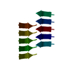
| ||||||||
| Unit cell |
|
-Components
| #1: Protein/peptide | Mass: 712.751 Da / Num. of mol.: 1 / Mutation: D290V / Source method: obtained synthetically / Source: (synth.) Homo sapiens (human) / References: UniProt: P22626 |
|---|---|
| #2: Water | ChemComp-HOH / Water Mass: 18.015 Da / Num. of mol.: 3 / Source method: isolated from a natural source / Formula: H2O Mass: 18.015 Da / Num. of mol.: 3 / Source method: isolated from a natural source / Formula: H2O |
-Experimental details
-Experiment
| Experiment | Method: X-RAY DIFFRACTION / Number of used crystals: 1 |
|---|
- Sample preparation
| Crystal grow | Temperature: 298 K / Method: vapor diffusion, hanging drop / pH: 5.5 Details: salt: 0.15M Ammonium Acetate, precipitant: 35% MPD, buffer: 0.1M Bis-Tris |
|---|
-Data collection
| Diffraction | Mean temperature: 100 K / Serial crystal experiment: N | ||||||||||||||||||||||||||||||||||||||||||||||||||||||||||||||||||||||||||||||||||||||||||||||||||||||||||||||||||||||||||||||||||||||||||||
|---|---|---|---|---|---|---|---|---|---|---|---|---|---|---|---|---|---|---|---|---|---|---|---|---|---|---|---|---|---|---|---|---|---|---|---|---|---|---|---|---|---|---|---|---|---|---|---|---|---|---|---|---|---|---|---|---|---|---|---|---|---|---|---|---|---|---|---|---|---|---|---|---|---|---|---|---|---|---|---|---|---|---|---|---|---|---|---|---|---|---|---|---|---|---|---|---|---|---|---|---|---|---|---|---|---|---|---|---|---|---|---|---|---|---|---|---|---|---|---|---|---|---|---|---|---|---|---|---|---|---|---|---|---|---|---|---|---|---|---|---|---|
| Diffraction source | Source: SYNCHROTRON / Site: APS / Beamline: 24-ID-E / Wavelength: 0.9792 Å | ||||||||||||||||||||||||||||||||||||||||||||||||||||||||||||||||||||||||||||||||||||||||||||||||||||||||||||||||||||||||||||||||||||||||||||
| Detector | Type: DECTRIS EIGER X 16M / Detector: PIXEL / Date: Nov 6, 2018 | ||||||||||||||||||||||||||||||||||||||||||||||||||||||||||||||||||||||||||||||||||||||||||||||||||||||||||||||||||||||||||||||||||||||||||||
| Radiation | Protocol: SINGLE WAVELENGTH / Monochromatic (M) / Laue (L): M / Scattering type: x-ray | ||||||||||||||||||||||||||||||||||||||||||||||||||||||||||||||||||||||||||||||||||||||||||||||||||||||||||||||||||||||||||||||||||||||||||||
| Radiation wavelength | Wavelength: 0.9792 Å / Relative weight: 1 | ||||||||||||||||||||||||||||||||||||||||||||||||||||||||||||||||||||||||||||||||||||||||||||||||||||||||||||||||||||||||||||||||||||||||||||
| Reflection | Resolution: 1.1→20.64 Å / Num. obs: 1329 / % possible obs: 86.5 % / Redundancy: 5.583 % / Biso Wilson estimate: 9.262 Å2 / CC1/2: 0.992 / Rmerge(I) obs: 0.106 / Rrim(I) all: 0.116 / Χ2: 0.918 / Net I/σ(I): 10.45 / Num. measured all: 7420 / Scaling rejects: 9 | ||||||||||||||||||||||||||||||||||||||||||||||||||||||||||||||||||||||||||||||||||||||||||||||||||||||||||||||||||||||||||||||||||||||||||||
| Reflection shell | Diffraction-ID: 1
|
- Processing
| Software |
| ||||||||||||||||||||||||||||||||||||||||||||||||||||||||||||||||||||||
|---|---|---|---|---|---|---|---|---|---|---|---|---|---|---|---|---|---|---|---|---|---|---|---|---|---|---|---|---|---|---|---|---|---|---|---|---|---|---|---|---|---|---|---|---|---|---|---|---|---|---|---|---|---|---|---|---|---|---|---|---|---|---|---|---|---|---|---|---|---|---|---|
| Refinement | Method to determine structure: AB INITIO PHASING / Resolution: 1.1→20.64 Å / Cor.coef. Fo:Fc: 0.992 / Cor.coef. Fo:Fc free: 0.99 / SU B: 0.676 / SU ML: 0.014 / SU R Cruickshank DPI: 0.0245 / Cross valid method: THROUGHOUT / σ(F): 0 / ESU R: 0.025 / ESU R Free: 0.026 Details: HYDROGENS HAVE BEEN USED IF PRESENT IN THE INPUT U VALUES : REFINED INDIVIDUALLY
| ||||||||||||||||||||||||||||||||||||||||||||||||||||||||||||||||||||||
| Solvent computation | Ion probe radii: 0.8 Å / Shrinkage radii: 0.8 Å / VDW probe radii: 1.2 Å | ||||||||||||||||||||||||||||||||||||||||||||||||||||||||||||||||||||||
| Displacement parameters | Biso max: 9.96 Å2 / Biso mean: 4.822 Å2 / Biso min: 3.84 Å2
| ||||||||||||||||||||||||||||||||||||||||||||||||||||||||||||||||||||||
| Refinement step | Cycle: final / Resolution: 1.1→20.64 Å
| ||||||||||||||||||||||||||||||||||||||||||||||||||||||||||||||||||||||
| Refine LS restraints |
| ||||||||||||||||||||||||||||||||||||||||||||||||||||||||||||||||||||||
| LS refinement shell | Resolution: 1.103→1.132 Å / Rfactor Rfree error: 0 / Total num. of bins used: 20
|
