 Movie
Movie Controller

[English] 日本語
 Yorodumi
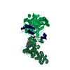
- PDB-6wo1: Hybrid acetohydroxyacid synthase complex structure with Cryptococ... -
+ Open data

- Basic information
| Entry | Database: PDB / ID: 6wo1 | ||||||
|---|---|---|---|---|---|---|---|
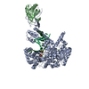
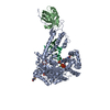
| Title | Hybrid acetohydroxyacid synthase complex structure with Cryptococcus neoformans AHAS catalytic subunit and Saccharomyces cerevisiae AHAS regulatory subunit | ||||||
 Components Components | (Acetohydroxyacid synthase ... Acetolactate synthase) x 2 Acetolactate synthase) x 2 | ||||||
 Keywords Keywords | TRANSFERASE / AHAS / pyruvate / FAD / dioxygen | ||||||
| Function / homology |  Function and homology information Function and homology informationacetolactate synthase regulator activity / acetolactate synthase complex / acetolactate synthase activity / acetolactate synthase / branched-chain amino acid biosynthetic process / valine biosynthetic process / isoleucine biosynthetic process / thiamine pyrophosphate binding / mitochondrial nucleoid / enzyme regulator activity ...acetolactate synthase regulator activity / acetolactate synthase complex / acetolactate synthase activity / acetolactate synthase / branched-chain amino acid biosynthetic process / valine biosynthetic process / isoleucine biosynthetic process / thiamine pyrophosphate binding / mitochondrial nucleoid / enzyme regulator activity / flavin adenine dinucleotide binding / magnesium ion binding / mitochondrionSimilarity search - Function | ||||||
| Biological species |  Cryptococcus neoformans (fungus) Cryptococcus neoformans (fungus) Saccharomyces cerevisiae (brewer's yeast) Saccharomyces cerevisiae (brewer's yeast) | ||||||
| Method | X-RAY DIFFRACTION / SYNCHROTRON / MOLECULAR REPLACEMENT / Resolution: 3.3 Å | ||||||
 Authors Authors | Guddat, L.W. / Lonhienne, T. | ||||||
| Funding support |  Australia, 1items Australia, 1items
| ||||||
 Citation Citation | Journal: Nature / Year: 2020 Title: Structures of fungal and plant acetohydroxyacid synthases. Authors: Thierry Lonhienne / Yu Shang Low / Mario D Garcia / Tristan Croll / Yan Gao / Quan Wang / Lou Brillault / Craig M Williams / James A Fraser / Ross P McGeary / Nicholas P West / Michael J ...Authors: Thierry Lonhienne / Yu Shang Low / Mario D Garcia / Tristan Croll / Yan Gao / Quan Wang / Lou Brillault / Craig M Williams / James A Fraser / Ross P McGeary / Nicholas P West / Michael J Landsberg / Zihe Rao / Gerhard Schenk / Luke W Guddat /   Abstract: Acetohydroxyacid synthase (AHAS), also known as acetolactate synthase, is a flavin adenine dinucleotide-, thiamine diphosphate- and magnesium-dependent enzyme that catalyses the first step in the ...Acetohydroxyacid synthase (AHAS), also known as acetolactate synthase, is a flavin adenine dinucleotide-, thiamine diphosphate- and magnesium-dependent enzyme that catalyses the first step in the biosynthesis of branched-chain amino acids. It is the target for more than 50 commercial herbicides. AHAS requires both catalytic and regulatory subunits for maximal activity and functionality. Here we describe structures of the hexadecameric AHAS complexes of Saccharomyces cerevisiae and dodecameric AHAS complexes of Arabidopsis thaliana. We found that the regulatory subunits of these AHAS complexes form a core to which the catalytic subunit dimers are attached, adopting the shape of a Maltese cross. The structures show how the catalytic and regulatory subunits communicate with each other to provide a pathway for activation and for feedback inhibition by branched-chain amino acids. We also show that the AHAS complex of Mycobacterium tuberculosis adopts a similar structure, thus demonstrating that the overall AHAS architecture is conserved across kingdoms. | ||||||
| History |
|
- Structure visualization
| Structure viewer | Molecule: MolmilJmol/JSmol |
|---|
- Downloads & links
-Download
| PDBx/mmCIF format | 6wo1.cif.gz | 310.7 KB | Display | PDBx/mmCIF format |
|---|---|---|---|---|
| PDB format | pdb6wo1.ent.gz | 249 KB | Display | PDB format |
| PDBx/mmJSON format | 6wo1.json.gz | Tree view | PDBx/mmJSON format | |
| Others |  Other downloads Other downloads |
-Validation report
| Arichive directory | https://data.pdbj.org/pub/pdb/validation_reports/wo/6wo1ftp://data.pdbj.org/pub/pdb/validation_reports/wo/6wo1 | HTTPS FTP |
|---|
-Related structure data
| Related structure data |  6u9dC  6u9hC  6vz8C  2fgcS 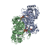 5imsS C: citing same article ( S: Starting model for refinement |
|---|---|
| Similar structure data |


-Links
 PDBj
PDBj


- Assembly
| Deposited unit | 
| ||||||||
|---|---|---|---|---|---|---|---|---|---|
| 1 |
| ||||||||
| Unit cell |
|
-Components
-Acetohydroxyacid synthase ... , 2 types, 2 molecules AB
| #1: Protein | Mass: 78594.672 Da / Num. of mol.: 1 Source method: isolated from a genetically manipulated source Source: (gene. exp.) Cryptococcus neoformans (fungus) / Production host:  Escherichia coli (E. coli) / References: UniProt: Q96VZ6, acetolactate synthase Escherichia coli (E. coli) / References: UniProt: Q96VZ6, acetolactate synthase |
|---|---|
| #2: Protein | Mass: 34041.734 Da / Num. of mol.: 1 Source method: isolated from a genetically manipulated source Source: (gene. exp.) Saccharomyces cerevisiae (brewer's yeast)Production host: Escherichia coli (E. coli) / References: UniProt: C7GQK9, UniProt: P25605*PLUS |
-Non-polymers , 5 types, 5 molecules 


| #3: Chemical | ChemComp-FAD / Flavin adenine dinucleotide Mass: 785.550 Da / Num. of mol.: 1 / Source method: obtained synthetically / Formula: C27H33N9O15P2 / Feature type: SUBJECT OF INVESTIGATION / Comment: FAD*YM Mass: 785.550 Da / Num. of mol.: 1 / Source method: obtained synthetically / Formula: C27H33N9O15P2 / Feature type: SUBJECT OF INVESTIGATION / Comment: FAD*YM |
|---|---|
| #4: Chemical | ChemComp-8GF /  Mass: 109.129 Da / Num. of mol.: 1 / Source method: obtained synthetically / Formula: C5H7N3 / Feature type: SUBJECT OF INVESTIGATION Mass: 109.129 Da / Num. of mol.: 1 / Source method: obtained synthetically / Formula: C5H7N3 / Feature type: SUBJECT OF INVESTIGATION |
| #5: Chemical | ChemComp-DPO / Pyrophosphate Mass: 173.943 Da / Num. of mol.: 1 / Source method: obtained synthetically / Formula: O7P2 / Feature type: SUBJECT OF INVESTIGATION Mass: 173.943 Da / Num. of mol.: 1 / Source method: obtained synthetically / Formula: O7P2 / Feature type: SUBJECT OF INVESTIGATION |
| #6: Chemical | ChemComp-MG /  Mass: 24.305 Da / Num. of mol.: 1 / Source method: isolated from a natural source / Formula: Mg / Feature type: SUBJECT OF INVESTIGATION Mass: 24.305 Da / Num. of mol.: 1 / Source method: isolated from a natural source / Formula: Mg / Feature type: SUBJECT OF INVESTIGATION |
| #7: Chemical | ChemComp-VAL / Valine Type: L-peptide linking / Mass: 117.146 Da / Num. of mol.: 1 / Source method: obtained synthetically / Formula: C5H11NO2 / Feature type: SUBJECT OF INVESTIGATION Type: L-peptide linking / Mass: 117.146 Da / Num. of mol.: 1 / Source method: obtained synthetically / Formula: C5H11NO2 / Feature type: SUBJECT OF INVESTIGATION |
-Details
| Has ligand of interest | Y |
|---|
-Experimental details
-Experiment
| Experiment | Method: X-RAY DIFFRACTION / Number of used crystals: 1 |
|---|
- Sample preparation
| Crystal | Density Matthews: 2.77 Å3/Da / Density % sol: 55.62 % |
|---|---|
| Crystal grow | Temperature: 291 K / Method: vapor diffusion, hanging drop / Details: Potassium thiocyanate, PEG 3350 |
-Data collection
| Diffraction | Mean temperature: 100 K / Ambient temp details: Oxford cryostream / Serial crystal experiment: N |
|---|---|
| Diffraction source | Source: SYNCHROTRON / Site: Australian Synchrotron / Beamline: MX1 / Wavelength: 0.9537 Å |
| Detector | Type: ADSC QUANTUM 210r / Detector: CCD / Date: Mar 28, 2019 |
| Radiation | Monochromator: SI(III) / Protocol: SINGLE WAVELENGTH / Monochromatic (M) / Laue (L): M / Scattering type: x-ray |
| Radiation wavelength | Wavelength: 0.9537 Å / Relative weight: 1 |
| Reflection | Resolution: 3.3→48.92 Å / Num. obs: 19890 / % possible obs: 99.8 % / Redundancy: 19.7 % / Biso Wilson estimate: 92.68 Å2 / CC1/2: 0.999 / Rpim(I) all: 0.041 / Net I/σ(I): 13.7 |
| Reflection shell | Resolution: 3.3→3.56 Å / Num. unique obs: 3985 / Rpim(I) all: 0.286 |
- Processing
| Software |
| ||||||||||||||||||||||||||||||||||||||||||||||||||||||||||||||||||||||||||||||||||||||||||||||||||||||||||||||||||||||||||||||||||||||||||||||||||||||||||||||||||||||||||||||||||||||||||||||||||||||||
|---|---|---|---|---|---|---|---|---|---|---|---|---|---|---|---|---|---|---|---|---|---|---|---|---|---|---|---|---|---|---|---|---|---|---|---|---|---|---|---|---|---|---|---|---|---|---|---|---|---|---|---|---|---|---|---|---|---|---|---|---|---|---|---|---|---|---|---|---|---|---|---|---|---|---|---|---|---|---|---|---|---|---|---|---|---|---|---|---|---|---|---|---|---|---|---|---|---|---|---|---|---|---|---|---|---|---|---|---|---|---|---|---|---|---|---|---|---|---|---|---|---|---|---|---|---|---|---|---|---|---|---|---|---|---|---|---|---|---|---|---|---|---|---|---|---|---|---|---|---|---|---|---|---|---|---|---|---|---|---|---|---|---|---|---|---|---|---|---|---|---|---|---|---|---|---|---|---|---|---|---|---|---|---|---|---|---|---|---|---|---|---|---|---|---|---|---|---|---|---|---|---|
| Refinement | Method to determine structure: MOLECULAR REPLACEMENT Starting model: 5IMS, 2FGC Resolution: 3.3→48.92 Å / SU ML: 0.42 / Cross valid method: THROUGHOUT / σ(F): 1.34 / Phase error: 26.7
| ||||||||||||||||||||||||||||||||||||||||||||||||||||||||||||||||||||||||||||||||||||||||||||||||||||||||||||||||||||||||||||||||||||||||||||||||||||||||||||||||||||||||||||||||||||||||||||||||||||||||
| Solvent computation | Shrinkage radii: 0.9 Å / VDW probe radii: 1.11 Å | ||||||||||||||||||||||||||||||||||||||||||||||||||||||||||||||||||||||||||||||||||||||||||||||||||||||||||||||||||||||||||||||||||||||||||||||||||||||||||||||||||||||||||||||||||||||||||||||||||||||||
| Displacement parameters | Biso max: 253.15 Å2 / Biso mean: 95.6873 Å2 / Biso min: 35.56 Å2 | ||||||||||||||||||||||||||||||||||||||||||||||||||||||||||||||||||||||||||||||||||||||||||||||||||||||||||||||||||||||||||||||||||||||||||||||||||||||||||||||||||||||||||||||||||||||||||||||||||||||||
| Refinement step | Cycle: final / Resolution: 3.3→48.92 Å
| ||||||||||||||||||||||||||||||||||||||||||||||||||||||||||||||||||||||||||||||||||||||||||||||||||||||||||||||||||||||||||||||||||||||||||||||||||||||||||||||||||||||||||||||||||||||||||||||||||||||||
| LS refinement shell | Refine-ID: X-RAY DIFFRACTION / Rfactor Rfree error: 0 / Total num. of bins used: 14
| ||||||||||||||||||||||||||||||||||||||||||||||||||||||||||||||||||||||||||||||||||||||||||||||||||||||||||||||||||||||||||||||||||||||||||||||||||||||||||||||||||||||||||||||||||||||||||||||||||||||||
| Refinement TLS params. | Method: refined / Refine-ID: X-RAY DIFFRACTION
| ||||||||||||||||||||||||||||||||||||||||||||||||||||||||||||||||||||||||||||||||||||||||||||||||||||||||||||||||||||||||||||||||||||||||||||||||||||||||||||||||||||||||||||||||||||||||||||||||||||||||
| Refinement TLS group |
|
