 Movie
Movie Controller

[English] 日本語
 Yorodumi
- PDB-6we7: HIV Integrase core domain in complex with inhibitor 3-methyl-2-{5... -
+ Open data

- Basic information
| Entry | Database: PDB / ID: 6we7 | ||||||
|---|---|---|---|---|---|---|---|
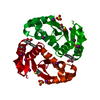
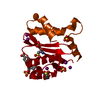
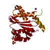
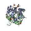
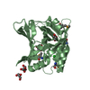
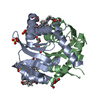
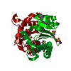
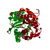
| Title | HIV Integrase core domain in complex with inhibitor 3-methyl-2-{5-methyl-2-[2-(thiophen-2-yl)ethynyl]-1- benzofuran-3-yl}butanoic acid | ||||||
 Components Components | Integrase | ||||||
 Keywords Keywords | TRANSFERASE/TRANSFERASE INHIBITOR / Inhibitor / HIV Integrase / TRANSFERASE-TRANSFERASE INHIBITOR complex | ||||||
| Function / homology |  Function and homology information Function and homology informationDNA integration / viral genome integration into host DNA / establishment of integrated proviral latency / RNA-directed DNA polymerase activity / endonuclease activity / DNA recombination / symbiont entry into host cell / DNA binding / zinc ion bindingSimilarity search - Function | ||||||
| Biological species |   Human immunodeficiency virus 1 Human immunodeficiency virus 1 | ||||||
| Method | X-RAY DIFFRACTION / SYNCHROTRON / FOURIER SYNTHESIS / Resolution: 2.28 Å | ||||||
 Authors Authors | Gorman, M.A. / Parker, M.W. | ||||||
 Citation Citation | Journal: To Be Published Title: HIV Integrase core domain in complex with inhibitor Authors: Gorman, M.A. / Parker, M.W. | ||||||
| History |
|
- Structure visualization
| Structure viewer | Molecule: MolmilJmol/JSmol |
|---|
- Downloads & links
-Download
| PDBx/mmCIF format | 6we7.cif.gz | 113.4 KB | Display | PDBx/mmCIF format |
|---|---|---|---|---|
| PDB format | pdb6we7.ent.gz | 93.1 KB | Display | PDB format |
| PDBx/mmJSON format | 6we7.json.gz | Tree view | PDBx/mmJSON format | |
| Others |  Other downloads Other downloads |
-Validation report
| Arichive directory | https://data.pdbj.org/pub/pdb/validation_reports/we/6we7ftp://data.pdbj.org/pub/pdb/validation_reports/we/6we7 | HTTPS FTP |
|---|
-Related structure data


-Links
 PDBj
PDBj- Assembly
| Deposited unit | 
| ||||||||
|---|---|---|---|---|---|---|---|---|---|
| 1 | 
| ||||||||
| Unit cell |
|
-Components
| #1: Protein | Mass: 18048.494 Da / Num. of mol.: 1 / Fragment: UNP residues 50-212 / Mutation: Q53E,C56S,G124S,A125T,W131E,V151I,F185K,Q209E Source method: isolated from a genetically manipulated source Source: (gene. exp.) Human immunodeficiency virus 1 / Gene: pol / Production host:  Escherichia coli (E. coli) Escherichia coli (E. coli)References: UniProt: F2WR52, Transferases; Transferring phosphorus-containing groups; Nucleotidyltransferases | ||||||||
|---|---|---|---|---|---|---|---|---|---|
| #2: Chemical | ChemComp-SO4 / Sulfate  Mass: 96.063 Da / Num. of mol.: 5 / Source method: obtained synthetically / Formula: SO4 Mass: 96.063 Da / Num. of mol.: 5 / Source method: obtained synthetically / Formula: SO4#3: Chemical | Iodide  Mass: 126.904 Da / Num. of mol.: 2 / Source method: obtained synthetically / Formula: I Mass: 126.904 Da / Num. of mol.: 2 / Source method: obtained synthetically / Formula: I#4: Chemical | ChemComp-TXM / ( |   Mass: 338.420 Da / Num. of mol.: 1 / Source method: obtained synthetically / Formula: C20H18O3S / Feature type: SUBJECT OF INVESTIGATION Mass: 338.420 Da / Num. of mol.: 1 / Source method: obtained synthetically / Formula: C20H18O3S / Feature type: SUBJECT OF INVESTIGATION#5: Water | ChemComp-HOH / | Water Mass: 18.015 Da / Num. of mol.: 34 / Source method: isolated from a natural source / Formula: H2O Mass: 18.015 Da / Num. of mol.: 34 / Source method: isolated from a natural source / Formula: H2OHas ligand of interest | Y | |
-Experimental details
-Experiment
| Experiment | Method: X-RAY DIFFRACTION / Number of used crystals: 1 |
|---|
- Sample preparation
| Crystal | Density Matthews: 2.07 Å3/Da / Density % sol: 40.48 % / Description: Bi-pyramid |
|---|---|
| Crystal grow | Temperature: 293 K / Method: vapor diffusion, hanging drop / Details: 2.2 M ammonium sulfate, 100 mM potassium iodide |
-Data collection
| Diffraction | Mean temperature: 100 K / Serial crystal experiment: N |
|---|---|
| Diffraction source | Source: SYNCHROTRON / Site: Australian Synchrotron  / Beamline: MX1 / Wavelength: 0.9537 Å / Beamline: MX1 / Wavelength: 0.9537 Å |
| Detector | Type: ADSC QUANTUM 210 / Detector: CCD / Date: May 16, 2015 |
| Radiation | Monochromator: Si(111) / Protocol: SINGLE WAVELENGTH / Monochromatic (M) / Laue (L): M / Scattering type: x-ray |
| Radiation wavelength | Wavelength: 0.9537 Å / Relative weight: 1 |
| Reflection | Resolution: 2.28→46.74 Å / Num. obs: 7524 / % possible obs: 99.7 % / Redundancy: 16.5 % / Biso Wilson estimate: 31.57 Å2 / CC1/2: 1 / Rmerge(I) obs: 0.079 / Rpim(I) all: 0.02 / Rrim(I) all: 0.082 / Χ2: 1.01 / Net I/σ(I): 34 |
| Reflection shell | Resolution: 2.28→2.35 Å / Redundancy: 16.7 % / Rmerge(I) obs: 0.703 / Mean I/σ(I) obs: 4.9 / Num. unique obs: 652 / CC1/2: 0.948 / Rpim(I) all: 0.174 / Rrim(I) all: 0.724 / Χ2: 1.04 / % possible all: 96.7 |
- Processing
| Software |
| ||||||||||||||||||||||||||||||||||||||||||||||||||||||||||||||||||||||||||||||||||||||||||||||||||||||||||||||||||||||||||||||||||||||||||||||||||||||||||||||||||||||||||||||||||||||
|---|---|---|---|---|---|---|---|---|---|---|---|---|---|---|---|---|---|---|---|---|---|---|---|---|---|---|---|---|---|---|---|---|---|---|---|---|---|---|---|---|---|---|---|---|---|---|---|---|---|---|---|---|---|---|---|---|---|---|---|---|---|---|---|---|---|---|---|---|---|---|---|---|---|---|---|---|---|---|---|---|---|---|---|---|---|---|---|---|---|---|---|---|---|---|---|---|---|---|---|---|---|---|---|---|---|---|---|---|---|---|---|---|---|---|---|---|---|---|---|---|---|---|---|---|---|---|---|---|---|---|---|---|---|---|---|---|---|---|---|---|---|---|---|---|---|---|---|---|---|---|---|---|---|---|---|---|---|---|---|---|---|---|---|---|---|---|---|---|---|---|---|---|---|---|---|---|---|---|---|---|---|---|---|
| Refinement | Method to determine structure: FOURIER SYNTHESIS / Resolution: 2.28→43.8 Å / Cor.coef. Fo:Fc: 0.937 / Cor.coef. Fo:Fc free: 0.885 / SU B: 14.235 / SU ML: 0.19 / Cross valid method: THROUGHOUT / ESU R: 0.359 / ESU R Free: 0.282 / Details: HYDROGENS HAVE BEEN ADDED IN THE RIDING POSITIONS
| ||||||||||||||||||||||||||||||||||||||||||||||||||||||||||||||||||||||||||||||||||||||||||||||||||||||||||||||||||||||||||||||||||||||||||||||||||||||||||||||||||||||||||||||||||||||
| Solvent computation | Ion probe radii: 0.8 Å / Shrinkage radii: 0.8 Å / VDW probe radii: 1.2 Å | ||||||||||||||||||||||||||||||||||||||||||||||||||||||||||||||||||||||||||||||||||||||||||||||||||||||||||||||||||||||||||||||||||||||||||||||||||||||||||||||||||||||||||||||||||||||
| Displacement parameters | Biso mean: 38.389 Å2
| ||||||||||||||||||||||||||||||||||||||||||||||||||||||||||||||||||||||||||||||||||||||||||||||||||||||||||||||||||||||||||||||||||||||||||||||||||||||||||||||||||||||||||||||||||||||
| Refinement step | Cycle: 1 / Resolution: 2.28→43.8 Å
| ||||||||||||||||||||||||||||||||||||||||||||||||||||||||||||||||||||||||||||||||||||||||||||||||||||||||||||||||||||||||||||||||||||||||||||||||||||||||||||||||||||||||||||||||||||||
| Refine LS restraints |
|