 Movie
Movie Controller

[English] 日本語
 Yorodumi
- PDB-6wbh: Crystal structure of mRECK(CC4) in fusion with engineered MBP at ... -
+ Open data

- Basic information
| Entry | Database: PDB / ID: 6wbh | ||||||||||||
|---|---|---|---|---|---|---|---|---|---|---|---|---|---|
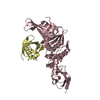
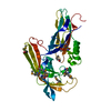
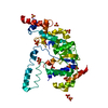
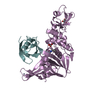
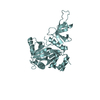
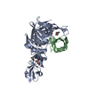
| Title | Crystal structure of mRECK(CC4) in fusion with engineered MBP at medium resolution | ||||||||||||
 Components Components | Maltodextrin-binding protein,Reversion-inducing cysteine-rich protein with Kazal motifs fusion | ||||||||||||
 Keywords Keywords |  SIGNALING PROTEIN / Wnt signaling / 4-helix bundle / extracellular domain / vascularization / blood-brain barrier / Maltose-binding protein SIGNALING PROTEIN / Wnt signaling / 4-helix bundle / extracellular domain / vascularization / blood-brain barrier / Maltose-binding protein | ||||||||||||
| Function / homology |  Function and homology information Function and homology informationregulation of establishment of blood-brain barrier / : / negative regulation of metalloendopeptidase activity / Post-translational modification: synthesis of GPI-anchored proteins / blood vessel maturation / : / metalloendopeptidase inhibitor activity / Wnt-protein binding / embryonic forelimb morphogenesis / Wnt signalosome ...regulation of establishment of blood-brain barrier / : / negative regulation of metalloendopeptidase activity / Post-translational modification: synthesis of GPI-anchored proteins / blood vessel maturation / : / metalloendopeptidase inhibitor activity / Wnt-protein binding / embryonic forelimb morphogenesis / Wnt signalosome / sprouting angiogenesis / endopeptidase inhibitor activity / detection of maltose stimulus / maltose transport complex / maltose binding / maltose transport / maltodextrin transmembrane transport / carbohydrate transport / carbohydrate transmembrane transporter activity / ATP-binding cassette (ABC) transporter complex, substrate-binding subunit-containing / regulation of angiogenesis / embryo implantation / ATP-binding cassette (ABC) transporter complex / extracellular matrix organization / cell chemotaxis / negative regulation of cell migration / serine-type endopeptidase inhibitor activity / positive regulation of canonical Wnt signaling pathway / outer membrane-bounded periplasmic space / periplasmic space / DNA damage response / membrane / plasma membraneSimilarity search - Function | ||||||||||||
| Biological species |  Escherichia coli (E. coli) Escherichia coli (E. coli) Mus musculus (house mouse) Mus musculus (house mouse) | ||||||||||||
| Method | X-RAY DIFFRACTION / MOLECULAR REPLACEMENT / molecular replacement / Resolution: 2.455 Å | ||||||||||||
| Model details | mouse RECK CC domain 4 in fusion with engineered Maltose Binding Protein at medium resolution | ||||||||||||
 Authors Authors | Chang, T.H. / Hsieh, F.L. / Gabelli, S.B. / Nathans, J. | ||||||||||||
| Funding support |  United States, United States,  France, 3items France, 3items
| ||||||||||||
 Citation Citation | Journal: Proc.Natl.Acad.Sci.USA / Year: 2020 Title: Structure of the RECK CC domain, an evolutionary anomaly. Authors: Chang, T.H. / Hsieh, F.L. / Smallwood, P.M. / Gabelli, S.B. / Nathans, J. | ||||||||||||
| History |
|
- Structure visualization
| Structure viewer | Molecule: MolmilJmol/JSmol |
|---|
- Downloads & links
-Download
| PDBx/mmCIF format | 6wbh.cif.gz | 256.4 KB | Display | PDBx/mmCIF format |
|---|---|---|---|---|
| PDB format | pdb6wbh.ent.gz | 207.5 KB | Display | PDB format |
| PDBx/mmJSON format | 6wbh.json.gz | Tree view | PDBx/mmJSON format | |
| Others |  Other downloads Other downloads |
-Validation report
| Arichive directory | https://data.pdbj.org/pub/pdb/validation_reports/wb/6wbhftp://data.pdbj.org/pub/pdb/validation_reports/wb/6wbh | HTTPS FTP |
|---|
-Related structure data
| Related structure data | 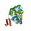 6wbjC 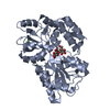 3setS S: Starting model for refinement C: citing same article ( |
|---|---|
| Similar structure data |

-Links
 PDBj
PDBj


- Assembly
| Deposited unit | 
| ||||||||
|---|---|---|---|---|---|---|---|---|---|
| 1 |
| ||||||||
| Unit cell |
|
-Components
| #1: Protein | Mass: 49096.473 Da / Num. of mol.: 1 Mutation: D84A,K85A,E174A,N175A,A217H,K221H,K241A,A314V,I319V,E361A,K364A,D365A Source method: isolated from a genetically manipulated source Source: (gene. exp.) Escherichia coli (E. coli), (gene. exp.) Mus musculus (house mouse)Gene: malE, DJ492_13065, EPS91_05465, FV295_14110, NCTC8450_00456, Reck Plasmid: pET11d / Production host: Escherichia coli (E. coli) / Strain (production host): SHuffle (DE3)References: UniProt: A0A376KDN7, UniProt: Q9Z0J1, UniProt: P0AEX9*PLUS | ||||
|---|---|---|---|---|---|
| #2: Polysaccharide | alpha-D-glucopyranose-(1-4)-alpha-D-glucopyranose / alpha-maltose  , Oligosaccharide / Class: Nutrient / Mass: 342.297 Da / Num. of mol.: 1 , Oligosaccharide / Class: Nutrient / Mass: 342.297 Da / Num. of mol.: 1Source method: isolated from a genetically manipulated source Details: oligosaccharide / References: alpha-maltose | ||||
| #3: Chemical | ChemComp-CL / Chloride  Mass: 35.453 Da / Num. of mol.: 1 / Source method: obtained synthetically / Formula: Cl / Feature type: SUBJECT OF INVESTIGATION Mass: 35.453 Da / Num. of mol.: 1 / Source method: obtained synthetically / Formula: Cl / Feature type: SUBJECT OF INVESTIGATION | ||||
| #4: Chemical |   Mass: 65.409 Da / Num. of mol.: 2 / Source method: obtained synthetically / Formula: Zn / Feature type: SUBJECT OF INVESTIGATION Mass: 65.409 Da / Num. of mol.: 2 / Source method: obtained synthetically / Formula: Zn / Feature type: SUBJECT OF INVESTIGATION#5: Water | ChemComp-HOH / | Water Mass: 18.015 Da / Num. of mol.: 218 / Source method: isolated from a natural source / Formula: H2O Mass: 18.015 Da / Num. of mol.: 218 / Source method: isolated from a natural source / Formula: H2OHas ligand of interest | Y | |
-Experimental details
-Experiment
| Experiment | Method: X-RAY DIFFRACTION / Number of used crystals: 1 |
|---|
- Sample preparation
| Crystal | Density Matthews: 2.61 Å3/Da / Density % sol: 52.89 % / Mosaicity: 0 ° |
|---|---|
| Crystal grow | Temperature: 293 K / Method: vapor diffusion / pH: 4.6 Details: 0.2 M ammonium sulfate, 0.1 M sodium acetate, pH 4.6, 25% PEG 4000 |
-Data collection
| Diffraction | Mean temperature: 100 K / Serial crystal experiment: N |
|---|---|
| Diffraction source | Source: ROTATING ANODE / Type: RIGAKU FR-E SUPERBRIGHT / Wavelength: 1.5419 Å |
| Detector | Type: DECTRIS PILATUS 200K / Detector: PIXEL / Date: Sep 10, 2018 |
| Radiation | Protocol: SINGLE WAVELENGTH / Monochromatic (M) / Laue (L): M / Scattering type: x-ray |
| Radiation wavelength | Wavelength: 1.5419 Å / Relative weight: 1 |
| Reflection | Resolution: 2.45→45.53 Å / Num. obs: 19054 / % possible obs: 98.3 % / Redundancy: 6.3 % / CC1/2: 0.947 / Rmerge(I) obs: 0.19 / Rpim(I) all: 0.081 / Rrim(I) all: 0.208 / Net I/σ(I): 7.4 |
| Reflection shell | Resolution: 2.45→2.51 Å / Redundancy: 5.7 % / Rmerge(I) obs: 0.511 / Num. unique obs: 1811 / CC1/2: 0.837 / Rpim(I) all: 0.222 / Rrim(I) all: 0.562 / % possible all: 94.52 |
-Phasing
| Phasing | Method: molecular replacement | |||||||||
|---|---|---|---|---|---|---|---|---|---|---|
| Phasing MR |
|
- Processing
| Software |
| |||||||||||||||||||||||||||||||||||||||||||||||||||||||||||||||||||||||||||||||||||||||||||||||||||||||||||||||||||||||||||||||||||||||||||||||||||||||||||||||||||||||||||||||
|---|---|---|---|---|---|---|---|---|---|---|---|---|---|---|---|---|---|---|---|---|---|---|---|---|---|---|---|---|---|---|---|---|---|---|---|---|---|---|---|---|---|---|---|---|---|---|---|---|---|---|---|---|---|---|---|---|---|---|---|---|---|---|---|---|---|---|---|---|---|---|---|---|---|---|---|---|---|---|---|---|---|---|---|---|---|---|---|---|---|---|---|---|---|---|---|---|---|---|---|---|---|---|---|---|---|---|---|---|---|---|---|---|---|---|---|---|---|---|---|---|---|---|---|---|---|---|---|---|---|---|---|---|---|---|---|---|---|---|---|---|---|---|---|---|---|---|---|---|---|---|---|---|---|---|---|---|---|---|---|---|---|---|---|---|---|---|---|---|---|---|---|---|---|---|---|---|
| Refinement | Method to determine structure: MOLECULAR REPLACEMENT Starting model: 3SET Resolution: 2.455→45.53 Å / SU ML: 0.27 / Cross valid method: THROUGHOUT / σ(F): 1.34 / Phase error: 19.12
| |||||||||||||||||||||||||||||||||||||||||||||||||||||||||||||||||||||||||||||||||||||||||||||||||||||||||||||||||||||||||||||||||||||||||||||||||||||||||||||||||||||||||||||||
| Solvent computation | Shrinkage radii: 0.9 Å / VDW probe radii: 1.11 Å | |||||||||||||||||||||||||||||||||||||||||||||||||||||||||||||||||||||||||||||||||||||||||||||||||||||||||||||||||||||||||||||||||||||||||||||||||||||||||||||||||||||||||||||||
| Displacement parameters | Biso max: 138.3 Å2 / Biso mean: 23.4561 Å2 / Biso min: 6.49 Å2 | |||||||||||||||||||||||||||||||||||||||||||||||||||||||||||||||||||||||||||||||||||||||||||||||||||||||||||||||||||||||||||||||||||||||||||||||||||||||||||||||||||||||||||||||
| Refinement step | Cycle: final / Resolution: 2.455→45.53 Å
| |||||||||||||||||||||||||||||||||||||||||||||||||||||||||||||||||||||||||||||||||||||||||||||||||||||||||||||||||||||||||||||||||||||||||||||||||||||||||||||||||||||||||||||||
| LS refinement shell | Refine-ID: X-RAY DIFFRACTION / Rfactor Rfree error: 0
| |||||||||||||||||||||||||||||||||||||||||||||||||||||||||||||||||||||||||||||||||||||||||||||||||||||||||||||||||||||||||||||||||||||||||||||||||||||||||||||||||||||||||||||||
| Refinement TLS params. | Method: refined / Refine-ID: X-RAY DIFFRACTION
| |||||||||||||||||||||||||||||||||||||||||||||||||||||||||||||||||||||||||||||||||||||||||||||||||||||||||||||||||||||||||||||||||||||||||||||||||||||||||||||||||||||||||||||||
| Refinement TLS group |
|
