 Movie
Movie Controller

[English] 日本語
 Yorodumi
- PDB-6w98: Single-Particle Cryo-EM Structure of Arabinofuranosyltransferase ... -
+ Open data

- Basic information
| Entry | Database: PDB / ID: 6w98 | |||||||||||||||||||||
|---|---|---|---|---|---|---|---|---|---|---|---|---|---|---|---|---|---|---|---|---|---|---|
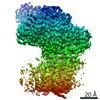
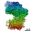
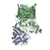
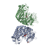
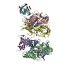
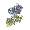
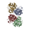
| Title | Single-Particle Cryo-EM Structure of Arabinofuranosyltransferase AftD from Mycobacteria | |||||||||||||||||||||
 Components Components |
| |||||||||||||||||||||
 Keywords Keywords |  MEMBRANE PROTEIN / Glycosyltransferase / lipomannan / lipoarabinomannan / arabinofuranose / nanodisc / single-particle cryo-electron microscopy / acyl carrier protein MEMBRANE PROTEIN / Glycosyltransferase / lipomannan / lipoarabinomannan / arabinofuranose / nanodisc / single-particle cryo-electron microscopy / acyl carrier protein | |||||||||||||||||||||
| Function / homology |  Function and homology informationacyl binding / lipid biosynthetic process / lipid A biosynthetic process / acyl carrier activity / phosphopantetheine binding / fatty acid biosynthetic process / transferase activity / membrane => GO:0016020 / response to xenobiotic stimulus / lipid binding ...acyl binding / lipid biosynthetic process / lipid A biosynthetic process / acyl carrier activity / phosphopantetheine binding / fatty acid biosynthetic process / transferase activity / membrane => GO:0016020 / response to xenobiotic stimulus / lipid binding / cytosol / cytoplasm Function and homology informationacyl binding / lipid biosynthetic process / lipid A biosynthetic process / acyl carrier activity / phosphopantetheine binding / fatty acid biosynthetic process / transferase activity / membrane => GO:0016020 / response to xenobiotic stimulus / lipid binding ...acyl binding / lipid biosynthetic process / lipid A biosynthetic process / acyl carrier activity / phosphopantetheine binding / fatty acid biosynthetic process / transferase activity / membrane => GO:0016020 / response to xenobiotic stimulus / lipid binding / cytosol / cytoplasmSimilarity search - Function | |||||||||||||||||||||
| Biological species |  Mycobacteroides abscessus subsp. abscessus (bacteria)Escherichia coli (E. coli) Mycobacteroides abscessus subsp. abscessus (bacteria)Escherichia coli (E. coli) | |||||||||||||||||||||
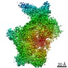
| Method | ELECTRON MICROSCOPY / single particle reconstruction / cryo EM / Resolution: 2.9 Å | |||||||||||||||||||||
 Authors Authors | Tan, Y.Z. / Zhang, L. / Rodrigues, J. / Zheng, R.B. / Giacometti, S.I. / Rosario, A.L. / Kloss, B. / Dandey, V.P. / Wei, H. / Brunton, R. ...Tan, Y.Z. / Zhang, L. / Rodrigues, J. / Zheng, R.B. / Giacometti, S.I. / Rosario, A.L. / Kloss, B. / Dandey, V.P. / Wei, H. / Brunton, R. / Raczkowski, A.M. / Athayde, D. / Catalao, M.J. / Pimentel, M. / Clarke, O.B. / Lowary, T.L. / Archer, M. / Niederweis, M. / Potter, C.S. / Carragher, B. / Mancia, F. | |||||||||||||||||||||
| Funding support |  United States, 6items United States, 6items
| |||||||||||||||||||||
 Citation Citation | Journal: Mol Cell / Year: 2020 Title: Cryo-EM Structures and Regulation of Arabinofuranosyltransferase AftD from Mycobacteria. Authors: Yong Zi Tan / Lei Zhang / José Rodrigues / Ruixiang Blake Zheng / Sabrina I Giacometti / Ana L Rosário / Brian Kloss / Venkata P Dandey / Hui Wei / Richard Brunton / Ashleigh M Raczkowski ...Authors: Yong Zi Tan / Lei Zhang / José Rodrigues / Ruixiang Blake Zheng / Sabrina I Giacometti / Ana L Rosário / Brian Kloss / Venkata P Dandey / Hui Wei / Richard Brunton / Ashleigh M Raczkowski / Diogo Athayde / Maria João Catalão / Madalena Pimentel / Oliver B Clarke / Todd L Lowary / Margarida Archer / Michael Niederweis / Clinton S Potter / Bridget Carragher / Filippo Mancia /    Abstract: Mycobacterium tuberculosis causes tuberculosis, a disease that kills over 1 million people each year. Its cell envelope is a common antibiotic target and has a unique structure due, in part, to two ...Mycobacterium tuberculosis causes tuberculosis, a disease that kills over 1 million people each year. Its cell envelope is a common antibiotic target and has a unique structure due, in part, to two lipidated polysaccharides-arabinogalactan and lipoarabinomannan. Arabinofuranosyltransferase D (AftD) is an essential enzyme involved in assembling these glycolipids. We present the 2.9-Å resolution structure of M. abscessus AftD, determined by single-particle cryo-electron microscopy. AftD has a conserved GT-C glycosyltransferase fold and three carbohydrate-binding modules. Glycan array analysis shows that AftD binds complex arabinose glycans. Additionally, AftD is non-covalently complexed with an acyl carrier protein (ACP). 3.4- and 3.5-Å structures of a mutant with impaired ACP binding reveal a conformational change, suggesting that ACP may regulate AftD function. Mutagenesis experiments using a conditional knockout constructed in M. smegmatis confirm the essentiality of the putative active site and the ACP binding for AftD function. | |||||||||||||||||||||
| History |
|
- Structure visualization
| Movie |
Movie viewer |
|---|---|
| Structure viewer | Molecule: MolmilJmol/JSmol |
- Downloads & links
-Download
| PDBx/mmCIF format | 6w98.cif.gz | 241.2 KB | Display | PDBx/mmCIF format |
|---|---|---|---|---|
| PDB format | pdb6w98.ent.gz | 192.8 KB | Display | PDB format |
| PDBx/mmJSON format | 6w98.json.gz | Tree view | PDBx/mmJSON format | |
| Others |  Other downloads Other downloads |
-Validation report
| Arichive directory | https://data.pdbj.org/pub/pdb/validation_reports/w9/6w98ftp://data.pdbj.org/pub/pdb/validation_reports/w9/6w98 | HTTPS FTP |
|---|
-Related structure data
| Related structure data | 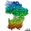 21580MC 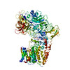 6wbxC 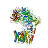 6wbyC M: map data used to model this data C: citing same article ( |
|---|---|
| Similar structure data | |
| EM raw data | EMPIAR-10399 (Title: Single-Particle Cryo-EM of Arabinofuranosyltransferase AftD from Mycobacteria, Wild-type Data size: 2.7 TB Data #1: Unaligned and compressed multi-frame movies [micrographs - multiframe] Data #2: Aligned and dose-weighted micrographs [micrographs - single frame] Data #3: Final Particle Stacks with Refined Euler Angles and Shifts (Overall and Focused Refined on CBM3) [picked particles - single frame - processed]) |


-Links
 PDBj
PDBj


- Assembly
| Deposited unit | 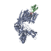
|
|---|---|
| 1 |
|
-Components
-Protein , 2 types, 2 molecules AB
| #1: Protein | Mass: 149584.281 Da / Num. of mol.: 1 Source method: isolated from a genetically manipulated source Source: (gene. exp.) Mycobacteroides abscessus subsp. abscessus (bacteria)Gene: SAMEA2161603_00457 / Production host: Escherichia coli (E. coli) / References: UniProt: A0A1N1NKH9 |
|---|---|
| #2: Protein | / ACP / Cytosolic-activating factor / CAF / Fatty acid synthase acyl carrier protein Mass: 8645.460 Da / Num. of mol.: 1 / Source method: isolated from a natural source / Source: (natural) Escherichia coli (strain K12) (bacteria) / Strain: K12 / References: UniProt: P0A6A8 |
-Non-polymers , 4 types, 5 molecules 

| #3: Chemical |  Mass: 40.078 Da / Num. of mol.: 2 / Source method: obtained synthetically / Formula: Ca / Feature type: SUBJECT OF INVESTIGATION Mass: 40.078 Da / Num. of mol.: 2 / Source method: obtained synthetically / Formula: Ca / Feature type: SUBJECT OF INVESTIGATION#4: Chemical | ChemComp-6OU / [( |  Mass: 717.996 Da / Num. of mol.: 1 / Source method: obtained synthetically / Formula: C39H76NO8P / Comment: phospholipid*YM Mass: 717.996 Da / Num. of mol.: 1 / Source method: obtained synthetically / Formula: C39H76NO8P / Comment: phospholipid*YM#5: Chemical | ChemComp-PNS / | Phosphopantetheine Mass: 358.348 Da / Num. of mol.: 1 / Source method: obtained synthetically / Formula: C11H23N2O7PS / Feature type: SUBJECT OF INVESTIGATION Mass: 358.348 Da / Num. of mol.: 1 / Source method: obtained synthetically / Formula: C11H23N2O7PS / Feature type: SUBJECT OF INVESTIGATION#6: Water | ChemComp-HOH / | WaterMass: 18.015 Da / Num. of mol.: 1 / Source method: isolated from a natural source / Formula: H2O |
|---|
-Details
| Has ligand of interest | Y |
|---|
-Experimental details
-Experiment
| Experiment | Method: ELECTRON MICROSCOPY |
|---|---|
| EM experiment | Aggregation state: PARTICLE / 3D reconstruction method: single particle reconstruction |
- Sample preparation
| Component |
| ||||||||||||||||||||||||
|---|---|---|---|---|---|---|---|---|---|---|---|---|---|---|---|---|---|---|---|---|---|---|---|---|---|
| Molecular weight |
| ||||||||||||||||||||||||
| Source (natural) |
| ||||||||||||||||||||||||
| Source (recombinant) | Organism: Escherichia coli (E. coli) | ||||||||||||||||||||||||
| Buffer solution | pH: 7.5 / Details: Solution was filtered and degassed. | ||||||||||||||||||||||||
| Buffer component |
| ||||||||||||||||||||||||
| Specimen | Conc.: 1 mg/ml / Embedding applied: NO / Shadowing applied: NO / Staining applied: NO / Vitrification applied: YES / Details: Protein was incorporated into lipid nanodiscs. | ||||||||||||||||||||||||
| Specimen support | Grid material: COPPER / Grid type: Homemade | ||||||||||||||||||||||||
| Vitrification | Instrument: SPOTITON / Cryogen name: ETHANE / Chamber temperature: 298 K Details: The Spotiton V1.0 robot was operating at room temperature and moderate humidity. |
- Electron microscopy imaging
| Experimental equipment |  Model: Titan Krios / Image courtesy: FEI Company |
|---|---|
| Microscopy | Model: FEI TITAN KRIOS |
| Electron gun | Electron source: FIELD EMISSION GUN / Accelerating voltage: 300 kV / Illumination mode: FLOOD BEAM |
| Electron lens | Mode: BRIGHT FIELDBright-field microscopy / Nominal magnification: 130000 X / Nominal defocus max: 2000 nm / Nominal defocus min: 1000 nm / Cs: 2.7 mm / Alignment procedure: ZEMLIN TABLEAU |
| Specimen holder | Cryogen: NITROGEN / Specimen holder model: FEI TITAN KRIOS AUTOGRID HOLDER |
| Image recording | Average exposure time: 13.05 sec. / Electron dose: 95.72 e/Å2 / Detector mode: COUNTING / Film or detector model: GATAN K2 QUANTUM (4k x 4k) / Num. of grids imaged: 4 / Num. of real images: 7274 |
| EM imaging optics | Energyfilter name: GIF Bioquantum / Energyfilter slit width: 15 eV |
| Image scans | Sampling size: 5 µm / Width: 3838 / Height: 3710 / Movie frames/image: 90 / Used frames/image: 1-90 |
- Processing
| EM software |
| ||||||||||||||||||||||||||||||||||||||||||||||||||||
|---|---|---|---|---|---|---|---|---|---|---|---|---|---|---|---|---|---|---|---|---|---|---|---|---|---|---|---|---|---|---|---|---|---|---|---|---|---|---|---|---|---|---|---|---|---|---|---|---|---|---|---|---|---|
| Image processing | Details: Energy filter slit width of 15 eV was used during the collection and was aligned automatically every hour using Leginon. | ||||||||||||||||||||||||||||||||||||||||||||||||||||
| CTF correction | Details: CTF was estimated using GCTF and refined throughout the pipeline using cisTEM. Type: PHASE FLIPPING AND AMPLITUDE CORRECTION | ||||||||||||||||||||||||||||||||||||||||||||||||||||
| Particle selection | Num. of particles selected: 2800102 | ||||||||||||||||||||||||||||||||||||||||||||||||||||
| Symmetry | Point symmetry: C1 (asymmetric) | ||||||||||||||||||||||||||||||||||||||||||||||||||||
| 3D reconstruction | Resolution: 2.9 Å / Resolution method: FSC 0.143 CUT-OFF / Num. of particles: 490616 / Symmetry type: POINT | ||||||||||||||||||||||||||||||||||||||||||||||||||||
| Atomic model building | Protocol: AB INITIO MODEL / Space: REAL |
