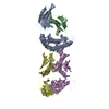
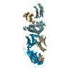
 Movie
Movie Controller


+ Open data

- Basic information
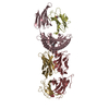
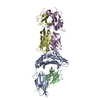
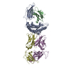
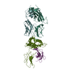
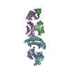
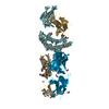
| Entry | Database: PDB / ID: 6vrm | ||||||
|---|---|---|---|---|---|---|---|
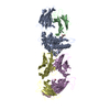
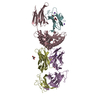
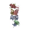
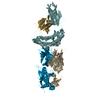
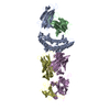
| Title | T cell receptor-p53-HLA-A2 complex | ||||||
 Components Components |
| ||||||
 Keywords Keywords |  IMMUNE SYSTEM / TCR complex / MHC / HLA / adoptive cell therapy IMMUNE SYSTEM / TCR complex / MHC / HLA / adoptive cell therapy | ||||||
| Function / homology |  Function and homology information Function and homology informationLoss of function of TP53 in cancer due to loss of tetramerization ability / Regulation of TP53 Expression / signal transduction by p53 class mediator / negative regulation of G1 to G0 transition / negative regulation of glucose catabolic process to lactate via pyruvate / Transcriptional activation of cell cycle inhibitor p21 / regulation of intrinsic apoptotic signaling pathway by p53 class mediator / Activation of NOXA and translocation to mitochondria / negative regulation of pentose-phosphate shunt / ATP-dependent DNA/DNA annealing activity ...Loss of function of TP53 in cancer due to loss of tetramerization ability / Regulation of TP53 Expression / signal transduction by p53 class mediator / negative regulation of G1 to G0 transition / negative regulation of glucose catabolic process to lactate via pyruvate / Transcriptional activation of cell cycle inhibitor p21 / regulation of intrinsic apoptotic signaling pathway by p53 class mediator / Activation of NOXA and translocation to mitochondria / negative regulation of pentose-phosphate shunt / ATP-dependent DNA/DNA annealing activity / negative regulation of helicase activity / regulation of cell cycle G2/M phase transition / intrinsic apoptotic signaling pathway in response to hypoxia / regulation of fibroblast apoptotic process / oxidative stress-induced premature senescence / oligodendrocyte apoptotic process / negative regulation of miRNA processing / positive regulation of thymocyte apoptotic process / glucose catabolic process to lactate via pyruvate / regulation of tissue remodeling / positive regulation of mitochondrial membrane permeability / negative regulation of mitophagy / positive regulation of programmed necrotic cell death / mRNA transcription / bone marrow development / circadian behavior / histone deacetylase regulator activity / regulation of mitochondrial membrane permeability involved in apoptotic process / RUNX3 regulates CDKN1A transcription / germ cell nucleus / regulation of DNA damage response, signal transduction by p53 class mediator / TP53 regulates transcription of additional cell cycle genes whose exact role in the p53 pathway remain uncertain / TP53 Regulates Transcription of Death Receptors and Ligands / Activation of PUMA and translocation to mitochondria / DNA damage response, signal transduction by p53 class mediator resulting in transcription of p21 class mediator / negative regulation of glial cell proliferation / Formation of Senescence-Associated Heterochromatin Foci (SAHF) / negative regulation of neuroblast proliferation / Regulation of TP53 Activity through Association with Co-factors / mitochondrial DNA repair / T cell lineage commitment / positive regulation of execution phase of apoptosis / negative regulation of DNA replication / ER overload response / B cell lineage commitment / thymocyte apoptotic process / TP53 regulates transcription of several additional cell death genes whose specific roles in p53-dependent apoptosis remain uncertain / positive regulation of cardiac muscle cell apoptotic process / TP53 Regulates Transcription of Caspase Activators and Caspases / T cell proliferation involved in immune response / cardiac septum morphogenesis / entrainment of circadian clock by photoperiod / PI5P Regulates TP53 Acetylation / Association of TriC/CCT with target proteins during biosynthesis / Zygotic genome activation (ZGA) / necroptotic process / rRNA transcription / positive regulation of release of cytochrome c from mitochondria / TP53 Regulates Transcription of Genes Involved in Cytochrome C Release / TFIID-class transcription factor complex binding / mitophagy / SUMOylation of transcription factors / negative regulation of telomere maintenance via telomerase / general transcription initiation factor binding / intrinsic apoptotic signaling pathway by p53 class mediator / antigen processing and presentation / Transcriptional Regulation by VENTX / response to X-ray / DNA damage response, signal transduction by p53 class mediator / replicative senescence / chromosome organization / neuroblast proliferation / intrinsic apoptotic signaling pathway in response to endoplasmic reticulum stress / cellular response to UV-C / : / hematopoietic stem cell differentiation / negative regulation of reactive oxygen species metabolic process / intrinsic apoptotic signaling pathway in response to DNA damage by p53 class mediator / glial cell proliferation / embryonic organ development / positive regulation of RNA polymerase II transcription preinitiation complex assembly / Pyroptosis / cis-regulatory region sequence-specific DNA binding / hematopoietic progenitor cell differentiation / cellular response to glucose starvation / cellular response to actinomycin D / TP53 Regulates Transcription of Genes Involved in G1 Cell Cycle Arrest / somitogenesis / type II interferon-mediated signaling pathway / positive regulation of intrinsic apoptotic signaling pathway / negative regulation of stem cell proliferation / core promoter sequence-specific DNA binding / gastrulation / negative regulation of fibroblast proliferation / MDM2/MDM4 family protein binding / cardiac muscle cell apoptotic process / transcription initiation-coupled chromatin remodeling / 14-3-3 protein binding / mitotic G1 DNA damage checkpoint signaling / Regulation of TP53 Activity through AcetylationSimilarity search - Function | ||||||
| Biological species |  Homo sapiens (human) Homo sapiens (human) | ||||||
| Method | X-RAY DIFFRACTION / SYNCHROTRON / MOLECULAR REPLACEMENT / Resolution: 2.61 Å | ||||||
 Authors Authors | Wu, D. / Gallagher, D.T. / Gowthaman, R. / Pierce, B.G. / Mariuzza, R.A. | ||||||
| Funding support |  United States, 1items United States, 1items
| ||||||
 Citation Citation | Journal: Nat Commun / Year: 2020 Title: Structural basis for oligoclonal T cell recognition of a shared p53 cancer neoantigen. Authors: Wu, D. / Gallagher, D.T. / Gowthaman, R. / Pierce, B.G. / Mariuzza, R.A. | ||||||
| History |
|
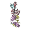
- Structure visualization
| Structure viewer | Molecule: MolmilJmol/JSmol |
|---|
- Downloads & links
-Download
| PDBx/mmCIF format | 6vrm.cif.gz | 335.1 KB | Display | PDBx/mmCIF format |
|---|---|---|---|---|
| PDB format | pdb6vrm.ent.gz | 271 KB | Display | PDB format |
| PDBx/mmJSON format | 6vrm.json.gz | Tree view | PDBx/mmJSON format | |
| Others |  Other downloads Other downloads |
-Validation report
| Arichive directory | https://data.pdbj.org/pub/pdb/validation_reports/vr/6vrmftp://data.pdbj.org/pub/pdb/validation_reports/vr/6vrm | HTTPS FTP |
|---|
-Related structure data
| Related structure data | 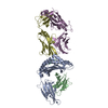 6vqoC  6vr1C  6vr5C  6vrnC  6vtcC  6vthC  2ak4S  3sjvS  5d2lS C: citing same article ( S: Starting model for refinement |
|---|---|
| Similar structure data |


-Links
 PDBj
PDBj


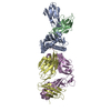
- Assembly
| Deposited unit | 
| ||||||||
|---|---|---|---|---|---|---|---|---|---|
| 1 |
| ||||||||
| Unit cell |
|
-Components
-Protein , 4 types, 4 molecules ABDE
| #1: Protein | Mass: 33912.469 Da / Num. of mol.: 1 Source method: isolated from a genetically manipulated source Source: (gene. exp.) Homo sapiens (human) / Gene: HLA-A / Production host:  Escherichia coli (E. coli) / References: UniProt: Q861F7, UniProt: A0A140T913*PLUS Escherichia coli (E. coli) / References: UniProt: Q861F7, UniProt: A0A140T913*PLUS |
|---|---|
| #2: Protein | Beta-2 microglobulin Mass: 11879.356 Da / Num. of mol.: 1 Source method: isolated from a genetically manipulated source Source: (gene. exp.) Homo sapiens (human) / Gene: B2M, CDABP0092, HDCMA22P / Production host: Escherichia coli (E. coli) / References: UniProt: P61769 |
| #3: Protein | Mass: 22881.312 Da / Num. of mol.: 1 Source method: isolated from a genetically manipulated source Source: (gene. exp.) Homo sapiens (human) / Production host: Escherichia coli (E. coli) |
| #4: Protein | Mass: 27789.844 Da / Num. of mol.: 1 Source method: isolated from a genetically manipulated source Source: (gene. exp.) Homo sapiens (human) / Production host: Escherichia coli (E. coli) |
-Protein/peptide / Non-polymers , 2 types, 22 molecules P
| #5: Protein/peptide | Mass: 1114.320 Da / Num. of mol.: 1 / Mutation: R175H Source method: isolated from a genetically manipulated source Source: (gene. exp.) Homo sapiens (human) / Gene: TP53, P53 / Production host: Escherichia coli (E. coli) / References: UniProt: P04637 |
|---|---|
| #6: Water | ChemComp-HOH / WaterMass: 18.015 Da / Num. of mol.: 21 / Source method: isolated from a natural source / Formula: H2O |
-Experimental details
-Experiment
| Experiment | Method: X-RAY DIFFRACTION / Number of used crystals: 1 |
|---|
- Sample preparation
| Crystal | Density Matthews: 3.14 Å3/Da / Density % sol: 60.8 % |
|---|---|
| Crystal grow | Temperature: 295 K / Method: vapor diffusion / pH: 7 Details: 20% (w/v) polyacrylic acid 5100, 0.1 M HEPES, 20 mM MgCl2 |
-Data collection
| Diffraction | Mean temperature: 100 K / Serial crystal experiment: N |
|---|---|
| Diffraction source | Source: SYNCHROTRON / Site: APS / Beamline: 23-ID-D / Wavelength: 1 Å |
| Detector | Type: DECTRIS PILATUS3 S 6M / Detector: PIXEL / Date: Jun 29, 2019 |
| Radiation | Protocol: SINGLE WAVELENGTH / Monochromatic (M) / Laue (L): M / Scattering type: x-ray |
| Radiation wavelength | Wavelength: 1 Å / Relative weight: 1 |
| Reflection | Resolution: 2.61→37.9 Å / Num. obs: 36118 / % possible obs: 98.4 % / Redundancy: 2.6 % / Rmerge(I) obs: 0.044 / Net I/σ(I): 12.2 |
| Reflection shell | Resolution: 2.61→2.7 Å / Rmerge(I) obs: 0.343 / Num. unique obs: 3641 |
- Processing
| Software |
| ||||||||||||||||||||||||||||||||||||||||||||||||||
|---|---|---|---|---|---|---|---|---|---|---|---|---|---|---|---|---|---|---|---|---|---|---|---|---|---|---|---|---|---|---|---|---|---|---|---|---|---|---|---|---|---|---|---|---|---|---|---|---|---|---|---|
| Refinement | Method to determine structure: MOLECULAR REPLACEMENT Starting model: 5D2L, 3SJV, 2AK4 Resolution: 2.61→19.79 Å / Cor.coef. Fo:Fc: 0.963 / Cor.coef. Fo:Fc free: 0.923 / SU B: 31.734 / SU ML: 0.289 / Cross valid method: THROUGHOUT / σ(F): 0 / ESU R Free: 0.316 / Stereochemistry target values: MAXIMUM LIKELIHOOD Details: HYDROGENS HAVE BEEN USED IF PRESENT IN THE INPUT U VALUES : REFINED INDIVIDUALLY
| ||||||||||||||||||||||||||||||||||||||||||||||||||
| Solvent computation | Ion probe radii: 0.8 Å / Shrinkage radii: 0.8 Å / VDW probe radii: 1.2 Å / Solvent model: MASK | ||||||||||||||||||||||||||||||||||||||||||||||||||
| Displacement parameters | Biso max: 144.86 Å2 / Biso mean: 75.679 Å2 / Biso min: 42.23 Å2
| ||||||||||||||||||||||||||||||||||||||||||||||||||
| Refinement step | Cycle: final / Resolution: 2.61→19.79 Å
| ||||||||||||||||||||||||||||||||||||||||||||||||||
| Refine LS restraints |
| ||||||||||||||||||||||||||||||||||||||||||||||||||
| LS refinement shell | Resolution: 2.61→2.677 Å / Rfactor Rfree error: 0 / Total num. of bins used: 20
|
