 Movie
Movie Controller


+ Open data

- Basic information
| Entry | Database: PDB / ID: 6vmt | ||||||
|---|---|---|---|---|---|---|---|
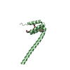
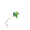
| Title | Leishmania major Programmed Cell Death Protein 5 Homolog | ||||||
 Components Components | Programmed Cell Death Protein 5 Homolog | ||||||
 Keywords Keywords | DNA BINDING PROTEIN / Structural Genomics / Structural Genomics of Pathogenic Protozoa Consortium / SGPP / PSI-2 / Protein Structure Initiative | ||||||
| Function / homology | PDCD5-like / PDCD5-like superfamily / Double-stranded DNA-binding domain / DNA binding / nucleus / cytosol / Uncharacterized protein Function and homology information Function and homology information | ||||||
| Biological species |  Leishmania major (eukaryote) Leishmania major (eukaryote) | ||||||
| Method | X-RAY DIFFRACTION / SYNCHROTRON / MOLECULAR REPLACEMENT / Resolution: 2.501 Å | ||||||
 Authors Authors | Merritt, E.A. / Structural Genomics of Pathogenic Protozoa Consortium (SGPP) | ||||||
 Citation Citation | Journal: To be published Title: Leishmania major Programmed Cell Death Protein 5 Homolog Authors: Merritt, E.A. / Anderson, L. / Arakaki, T. / Hol, W.G. / Le Trong, I. / Mehlin, C. / Soltis, M. / Structural Genomics of Pathogenic Protozoa Consortium (SGPP) | ||||||
| History |
|
- Structure visualization
| Structure viewer | Molecule: MolmilJmol/JSmol |
|---|
- Downloads & links
-Download
| PDBx/mmCIF format | 6vmt.cif.gz | 35.1 KB | Display | PDBx/mmCIF format |
|---|---|---|---|---|
| PDB format | pdb6vmt.ent.gz | 21.3 KB | Display | PDB format |
| PDBx/mmJSON format | 6vmt.json.gz | Tree view | PDBx/mmJSON format | |
| Others |  Other downloads Other downloads |
-Validation report
| Arichive directory | https://data.pdbj.org/pub/pdb/validation_reports/vm/6vmtftp://data.pdbj.org/pub/pdb/validation_reports/vm/6vmt | HTTPS FTP |
|---|
-Related structure data
| Related structure data |  6iqoS S: Starting model for refinement |
|---|---|
| Similar structure data | |
| Other databases |


-Links
 PDBj
PDBj- Assembly
| Deposited unit | 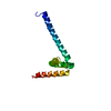
| ||||||||
|---|---|---|---|---|---|---|---|---|---|
| 1 |
| ||||||||
| Unit cell |
|
-Components
| #1: Protein | Mass: 13304.014 Da / Num. of mol.: 1 Source method: isolated from a genetically manipulated source Source: (gene. exp.) Leishmania major (eukaryote) / Gene: LMJF_28_1920 / Production host:  Escherichia coli (E. coli) / References: UniProt: Q4Q874 Escherichia coli (E. coli) / References: UniProt: Q4Q874 |
|---|---|
| #2: Water | ChemComp-HOH / Water Mass: 18.015 Da / Num. of mol.: 16 / Source method: isolated from a natural source / Formula: H2O Mass: 18.015 Da / Num. of mol.: 16 / Source method: isolated from a natural source / Formula: H2O |
-Experimental details
-Experiment
| Experiment | Method: X-RAY DIFFRACTION / Number of used crystals: 1 |
|---|
- Sample preparation
| Crystal | Density Matthews: 2.82 Å3/Da / Density % sol: 56.43 % |
|---|---|
| Crystal grow | Temperature: 300 K / Method: vapor diffusion, sitting drop / pH: 6.6 / Details: 1.5 M K2HPO4 |
-Data collection
| Diffraction | Mean temperature: 100 K / Serial crystal experiment: N | ||||||||||||||||||||||||
|---|---|---|---|---|---|---|---|---|---|---|---|---|---|---|---|---|---|---|---|---|---|---|---|---|---|
| Diffraction source | Source: SYNCHROTRON / Site: ALS  / Beamline: 8.2.2 / Wavelength: 0.95373,0.97962,0.97949 / Beamline: 8.2.2 / Wavelength: 0.95373,0.97962,0.97949 | ||||||||||||||||||||||||
| Detector | Type: ADSC QUANTUM 315 / Detector: CCD / Date: Mar 11, 2005 | ||||||||||||||||||||||||
| Radiation | Protocol: MAD / Monochromatic (M) / Laue (L): M / Scattering type: x-ray | ||||||||||||||||||||||||
| Radiation wavelength |
| ||||||||||||||||||||||||
| Reflection | Resolution: 2.5→25.296 Å / Num. obs: 4643 / % possible obs: 87.8 % / Redundancy: 8.9 % / CC1/2: 0.998 / Rmerge(I) obs: 0.114 / Rpim(I) all: 0.04 / Rrim(I) all: 0.122 / Net I/σ(I): 11.3 | ||||||||||||||||||||||||
| Reflection shell | Diffraction-ID: 1
|
- Processing
| Software |
| ||||||||||||||||||||||||||||||||||||||||||||||||||||||||||||||||||||||||||||||||||||||||||||||||||||||||||||||||||||||||||||||||||||||||||||||||||||||
|---|---|---|---|---|---|---|---|---|---|---|---|---|---|---|---|---|---|---|---|---|---|---|---|---|---|---|---|---|---|---|---|---|---|---|---|---|---|---|---|---|---|---|---|---|---|---|---|---|---|---|---|---|---|---|---|---|---|---|---|---|---|---|---|---|---|---|---|---|---|---|---|---|---|---|---|---|---|---|---|---|---|---|---|---|---|---|---|---|---|---|---|---|---|---|---|---|---|---|---|---|---|---|---|---|---|---|---|---|---|---|---|---|---|---|---|---|---|---|---|---|---|---|---|---|---|---|---|---|---|---|---|---|---|---|---|---|---|---|---|---|---|---|---|---|---|---|---|---|---|---|---|
| Refinement | Method to determine structure: MOLECULAR REPLACEMENT Starting model: 6IQO Resolution: 2.501→25.296 Å / Cor.coef. Fo:Fc: 0.951 / Cor.coef. Fo:Fc free: 0.919 / SU B: 11.938 / SU ML: 0.242 / Cross valid method: FREE R-VALUE / ESU R: 0.471 / ESU R Free: 0.306 Details: Hydrogens have been added in their riding positions. Sulfur f'' term set to 2.5e to account for partial substitution of SeMet for Met
| ||||||||||||||||||||||||||||||||||||||||||||||||||||||||||||||||||||||||||||||||||||||||||||||||||||||||||||||||||||||||||||||||||||||||||||||||||||||
| Solvent computation | Ion probe radii: 0.8 Å / Shrinkage radii: 0.8 Å / VDW probe radii: 1.4 Å | ||||||||||||||||||||||||||||||||||||||||||||||||||||||||||||||||||||||||||||||||||||||||||||||||||||||||||||||||||||||||||||||||||||||||||||||||||||||
| Displacement parameters | Biso mean: 79.593 Å2
| ||||||||||||||||||||||||||||||||||||||||||||||||||||||||||||||||||||||||||||||||||||||||||||||||||||||||||||||||||||||||||||||||||||||||||||||||||||||
| Refinement step | Cycle: LAST / Resolution: 2.501→25.296 Å
| ||||||||||||||||||||||||||||||||||||||||||||||||||||||||||||||||||||||||||||||||||||||||||||||||||||||||||||||||||||||||||||||||||||||||||||||||||||||
| Refine LS restraints |
| ||||||||||||||||||||||||||||||||||||||||||||||||||||||||||||||||||||||||||||||||||||||||||||||||||||||||||||||||||||||||||||||||||||||||||||||||||||||
| LS refinement shell |
|
