 Movie
Movie Controller

[English] 日本語
 Yorodumi
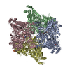
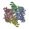
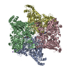
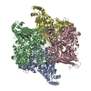
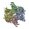
- PDB-6vl7: Crystal structure of the H583C mutant of GoxA soaked with glycine -
+ Open data

- Basic information
| Entry | Database: PDB / ID: 6vl7 | ||||||
|---|---|---|---|---|---|---|---|
| Title | Crystal structure of the H583C mutant of GoxA soaked with glycine | ||||||
 Components Components | Glycine oxidase | ||||||
 Keywords Keywords | OXIDOREDUCTASE / CTQ | ||||||
| Function / homology | L-Lysine epsilon oxidase, N-terminal / L-lysine epsilon oxidase, C-terminal / L-Lysine epsilon oxidase N-terminal / L-lysine epsilon oxidase C-terminal domain / DI(HYDROXYETHYL)ETHER / TRIETHYLENE GLYCOL / Uncharacterized protein Function and homology information Function and homology information | ||||||
| Biological species |  Pseudoalteromonas luteoviolacea DSM 6061 (bacteria) Pseudoalteromonas luteoviolacea DSM 6061 (bacteria) | ||||||
| Method | X-RAY DIFFRACTION / SYNCHROTRON / FOURIER SYNTHESIS / Resolution: 2.14 Å | ||||||
 Authors Authors | Yukl, E.T. | ||||||
 Citation Citation | Journal: J.Biol.Chem. / Year: 2020 Title: Roles of active-site residues in catalysis, substrate binding, cooperativity, and the reaction mechanism of the quinoprotein glycine oxidase. Authors: Mamounis, K.J. / Yukl, E.T. / Davidson, V.L. | ||||||
| History |
|
- Structure visualization
| Structure viewer | Molecule: MolmilJmol/JSmol |
|---|
- Downloads & links
-Download
| PDBx/mmCIF format | 6vl7.cif.gz | 1.3 MB | Display | PDBx/mmCIF format |
|---|---|---|---|---|
| PDB format | pdb6vl7.ent.gz | 1.1 MB | Display | PDB format |
| PDBx/mmJSON format | 6vl7.json.gz | Tree view | PDBx/mmJSON format | |
| Others |  Other downloads Other downloads |
-Validation report
| Arichive directory | https://data.pdbj.org/pub/pdb/validation_reports/vl/6vl7ftp://data.pdbj.org/pub/pdb/validation_reports/vl/6vl7 | HTTPS FTP |
|---|
-Related structure data
| Related structure data |  6vmfC 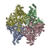 6vmvC  6vmwC  6bywS S: Starting model for refinement C: citing same article ( |
|---|---|
| Similar structure data |


-Links
 PDBj
PDBj- Assembly
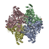
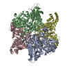
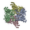
| Deposited unit | 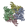
| ||||||||
|---|---|---|---|---|---|---|---|---|---|
| 1 |
| ||||||||
| Unit cell |
|
-Components
-Protein , 1 types, 4 molecules ABCD
| #1: Protein | Mass: 91491.781 Da / Num. of mol.: 4 / Mutation: H583C Source method: isolated from a genetically manipulated source Source: (gene. exp.) Pseudoalteromonas luteoviolacea DSM 6061 (bacteria)Gene: N475_19905 / Production host: Escherichia coli (E. coli) / References: UniProt: A0A161XU12 |
|---|
-Non-polymers , 5 types, 1562 molecules 


| #2: Chemical | ChemComp-MG /  Mass: 24.305 Da / Num. of mol.: 4 / Source method: obtained synthetically / Formula: Mg Mass: 24.305 Da / Num. of mol.: 4 / Source method: obtained synthetically / Formula: Mg#3: Chemical | ChemComp-SO4 / Sulfate Mass: 96.063 Da / Num. of mol.: 10 / Source method: obtained synthetically / Formula: SO4 Mass: 96.063 Da / Num. of mol.: 10 / Source method: obtained synthetically / Formula: SO4#4: Chemical | ChemComp-PEG / | Diethylene glycol Mass: 106.120 Da / Num. of mol.: 1 / Source method: obtained synthetically / Formula: C4H10O3 Mass: 106.120 Da / Num. of mol.: 1 / Source method: obtained synthetically / Formula: C4H10O3#5: Chemical | Polyethylene glycol Mass: 150.173 Da / Num. of mol.: 3 / Source method: obtained synthetically / Formula: C6H14O4 Mass: 150.173 Da / Num. of mol.: 3 / Source method: obtained synthetically / Formula: C6H14O4#6: Water | ChemComp-HOH / | WaterMass: 18.015 Da / Num. of mol.: 1544 / Source method: isolated from a natural source / Formula: H2O |
|---|
-Details
| Has ligand of interest | N |
|---|
-Experimental details
-Experiment
| Experiment | Method: X-RAY DIFFRACTION / Number of used crystals: 1 |
|---|
- Sample preparation
| Crystal | Density Matthews: 2.63 Å3/Da / Density % sol: 53.21 % |
|---|---|
| Crystal grow | Temperature: 293 K / Method: batch mode / pH: 7.5 Details: 1uL protein was combined with 1uL precipitant solution containing 25% PEG 3350, 0.1 M HEPES pH 7.5 and 0.1 M ammonium sulfate |
-Data collection
| Diffraction | Mean temperature: 100 K / Serial crystal experiment: N |
|---|---|
| Diffraction source | Source: SYNCHROTRON / Site: ALS  / Beamline: 5.0.2 / Wavelength: 1 Å / Beamline: 5.0.2 / Wavelength: 1 Å |
| Detector | Type: DECTRIS PILATUS 6M / Detector: PIXEL / Date: Jun 30, 2018 |
| Radiation | Monochromator: Double-crystal, Si(111) / Protocol: SINGLE WAVELENGTH / Monochromatic (M) / Laue (L): M / Scattering type: x-ray |
| Radiation wavelength | Wavelength: 1 Å / Relative weight: 1 |
| Reflection | Resolution: 2.14→49.191 Å / Num. obs: 207973 / % possible obs: 99.4 % / Redundancy: 3.7 % / CC1/2: 0.997 / Rmerge(I) obs: 0.087 / Rpim(I) all: 0.051 / Rrim(I) all: 0.101 / Net I/σ(I): 9.2 |
| Reflection shell | Resolution: 2.14→2.18 Å / Redundancy: 3.9 % / Rmerge(I) obs: 0.605 / Mean I/σ(I) obs: 2.1 / Num. unique obs: 10307 / CC1/2: 0.783 / Rpim(I) all: 0.354 / Rrim(I) all: 0.703 / % possible all: 100 |
- Processing
| Software |
| ||||||||||||||||||||||||||||||||||||||||||||||||||||||||||||||||||||||||||||||||||||||||||||||||||||||||||||
|---|---|---|---|---|---|---|---|---|---|---|---|---|---|---|---|---|---|---|---|---|---|---|---|---|---|---|---|---|---|---|---|---|---|---|---|---|---|---|---|---|---|---|---|---|---|---|---|---|---|---|---|---|---|---|---|---|---|---|---|---|---|---|---|---|---|---|---|---|---|---|---|---|---|---|---|---|---|---|---|---|---|---|---|---|---|---|---|---|---|---|---|---|---|---|---|---|---|---|---|---|---|---|---|---|---|---|---|---|---|
| Refinement | Method to determine structure: FOURIER SYNTHESIS Starting model: 6BYW Resolution: 2.14→49.191 Å / SU ML: 0.22 / Cross valid method: THROUGHOUT / σ(F): 1.33 / Phase error: 23.73
| ||||||||||||||||||||||||||||||||||||||||||||||||||||||||||||||||||||||||||||||||||||||||||||||||||||||||||||
| Solvent computation | Shrinkage radii: 0.9 Å / VDW probe radii: 1.11 Å | ||||||||||||||||||||||||||||||||||||||||||||||||||||||||||||||||||||||||||||||||||||||||||||||||||||||||||||
| Displacement parameters | Biso max: 162.47 Å2 / Biso mean: 47.6067 Å2 / Biso min: 18.09 Å2 | ||||||||||||||||||||||||||||||||||||||||||||||||||||||||||||||||||||||||||||||||||||||||||||||||||||||||||||
| Refinement step | Cycle: final / Resolution: 2.14→49.191 Å
| ||||||||||||||||||||||||||||||||||||||||||||||||||||||||||||||||||||||||||||||||||||||||||||||||||||||||||||
| LS refinement shell | Refine-ID: X-RAY DIFFRACTION / Rfactor Rfree error: 0
| ||||||||||||||||||||||||||||||||||||||||||||||||||||||||||||||||||||||||||||||||||||||||||||||||||||||||||||
| Refinement TLS params. | Method: refined / Origin x: 16.1741 Å / Origin y: -142.5305 Å / Origin z: 233.4471 Å
| ||||||||||||||||||||||||||||||||||||||||||||||||||||||||||||||||||||||||||||||||||||||||||||||||||||||||||||
| Refinement TLS group |
|
