 Movie
Movie Controller

[English] 日本語
 Yorodumi
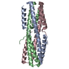
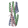
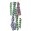
- PDB-6vjo: Human parainfluenza virus type 3 fusion glycoprotein N-terminal h... -
+ Open data

- Basic information
| Entry | Database: PDB / ID: 6vjo | |||||||||
|---|---|---|---|---|---|---|---|---|---|---|
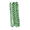
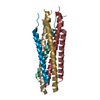
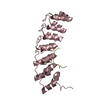
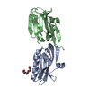
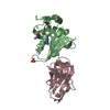
| Title | Human parainfluenza virus type 3 fusion glycoprotein N-terminal heptad repeat domain+alpha/beta-VI | |||||||||
 Components Components | (Fusion glycoprotein F0) x 2 | |||||||||
 Keywords Keywords |  ANTIVIRAL PROTEIN / Fusion protein / fusion inhibitor / six-helix bundle / alpha/beta-peptide ANTIVIRAL PROTEIN / Fusion protein / fusion inhibitor / six-helix bundle / alpha/beta-peptide | |||||||||
| Function / homology |  Function and homology information Function and homology informationmembrane => GO:0016020 / fusion of virus membrane with host plasma membrane / viral envelope / host cell plasma membrane / virion membrane / plasma membraneSimilarity search - Function | |||||||||
| Biological species |  Human respirovirus 3 Human respirovirus 3 | |||||||||
| Method | X-RAY DIFFRACTION / MOLECULAR REPLACEMENT / Resolution: 2 Å | |||||||||
 Authors Authors | Outlaw, V.K. / Kreitler, D.F. / Gellman, S.H. | |||||||||
| Funding support |  United States, 2items United States, 2items
| |||||||||
 Citation Citation | Journal: J.Am.Chem.Soc. / Year: 2021 Title: Engineering Protease-Resistant Peptides to Inhibit Human Parainfluenza Viral Respiratory Infection. Authors: Outlaw, V.K. / Cheloha, R.W. / Jurgens, E.M. / Bovier, F.T. / Zhu, Y. / Kreitler, D.F. / Harder, O. / Niewiesk, S. / Porotto, M. / Gellman, S.H. / Moscona, A. | |||||||||
| History |
|
- Structure visualization
| Structure viewer | Molecule: MolmilJmol/JSmol |
|---|
- Downloads & links
-Download
| PDBx/mmCIF format | 6vjo.cif.gz | 77.5 KB | Display | PDBx/mmCIF format |
|---|---|---|---|---|
| PDB format | pdb6vjo.ent.gz | 49.4 KB | Display | PDB format |
| PDBx/mmJSON format | 6vjo.json.gz | Tree view | PDBx/mmJSON format | |
| Others |  Other downloads Other downloads |
-Validation report
| Arichive directory | https://data.pdbj.org/pub/pdb/validation_reports/vj/6vjoftp://data.pdbj.org/pub/pdb/validation_reports/vj/6vjo | HTTPS FTP |
|---|
-Related structure data
| Related structure data | 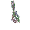 1ztmS S: Starting model for refinement |
|---|---|
| Similar structure data |

-Links
 PDBj
PDBj

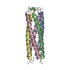
- Assembly
| Deposited unit | 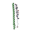
| ||||||||||||
|---|---|---|---|---|---|---|---|---|---|---|---|---|---|
| 1 | 
| ||||||||||||
| Unit cell |
|
-Components
| #1: Protein/peptide | Mass: 4191.891 Da / Num. of mol.: 1 Mutation: I458(XCP), E459V, K462(XPC), A463I, S465(XCP), E469(B3E), E472(B3E), R476(XPC), Q479(XCP), S483(XCP) Source method: obtained synthetically Details: alpha/beta-VI is a synthetic alpha/beta peptide derived from residues 449-484 of the HPIV3 fusion glycoprotein C-terminal heptad repeat domain with substitutions I458(XCP), E459V, K462(XPC), ...Details: alpha/beta-VI is a synthetic alpha/beta peptide derived from residues 449-484 of the HPIV3 fusion glycoprotein C-terminal heptad repeat domain with substitutions I458(XCP), E459V, K462(XPC), A463I, S465(XCP), E469(B3E), E472(B3E), R476(XPC), Q479(XCP), S483(XCP). It is acetylated at the N-terminus and amidated at the C-terminus. Source: (synth.) Human respirovirus 3 / References: UniProt: A0A1X9QNY3 |
|---|---|
| #2: Protein | Mass: 5656.473 Da / Num. of mol.: 1 / Source method: obtained synthetically Details: This compound is derived from residues 139-189 of the HPIV3 fusion glycoprotein. It is acetylated at the N-terminus and amidated at the C-terminus. Source: (synth.) Human respirovirus 3 / References: UniProt: Q84193 |
| #3: Water | ChemComp-HOH / Water Mass: 18.015 Da / Num. of mol.: 35 / Source method: isolated from a natural source / Formula: H2O Mass: 18.015 Da / Num. of mol.: 35 / Source method: isolated from a natural source / Formula: H2O |
| Has ligand of interest | N |
-Experimental details
-Experiment
| Experiment | Method: X-RAY DIFFRACTION / Number of used crystals: 1 |
|---|
- Sample preparation
| Crystal | Density Matthews: 3.07 Å3/Da / Density % sol: 59.97 % |
|---|---|
| Crystal grow | Temperature: 298 K / Method: vapor diffusion, hanging drop / pH: 6.5 Details: 30 mM NaF, 30 mM NaBr, 30 mM NaI, 20% (v/v) PEG 500 MME, 10% (w/v) PEG 20000 in 100 mM imidazole/MES monohydrate buffer (pH 6.5) |
-Data collection
| Diffraction | Mean temperature: 100 K / Serial crystal experiment: N |
|---|---|
| Diffraction source | Source: ROTATING ANODE / Type: BRUKER AXS MICROSTAR / Wavelength: 1.542 Å |
| Detector | Type: BRUKER SMART 6000 / Detector: CCD / Date: Jul 1, 2016 |
| Radiation | Protocol: SINGLE WAVELENGTH / Monochromatic (M) / Laue (L): M / Scattering type: x-ray |
| Radiation wavelength | Wavelength: 1.542 Å / Relative weight: 1 |
| Reflection | Resolution: 2→35.55 Å / Num. obs: 7675 / % possible obs: 96.8 % / Redundancy: 8.7 % / Biso Wilson estimate: 16.48 Å2 / CC1/2: 0.996 / Rpim(I) all: 0.058 / Rrim(I) all: 0.171 / Rsym value: 0.161 / Net I/σ(I): 10.5 |
| Reflection shell | Resolution: 2→2.13 Å / Mean I/σ(I) obs: 2.4 / Num. unique obs: 1107 / CC1/2: 0.426 / Rpim(I) all: 0.478 / Rrim(I) all: 0.944 / Rsym value: 0.161 / % possible all: 83.9 |
- Processing
| Software |
| |||||||||||||||||||||||||||||||||||||||||||||||||
|---|---|---|---|---|---|---|---|---|---|---|---|---|---|---|---|---|---|---|---|---|---|---|---|---|---|---|---|---|---|---|---|---|---|---|---|---|---|---|---|---|---|---|---|---|---|---|---|---|---|---|
| Refinement | Method to determine structure: MOLECULAR REPLACEMENT Starting model: 1ZTM Resolution: 2→35.55 Å / SU ML: 0.3378 / Cross valid method: FREE R-VALUE / σ(F): 2 / Phase error: 26.9055
| |||||||||||||||||||||||||||||||||||||||||||||||||
| Solvent computation | Shrinkage radii: 0.9 Å / VDW probe radii: 1.11 Å | |||||||||||||||||||||||||||||||||||||||||||||||||
| Displacement parameters | Biso mean: 32.3 Å2 | |||||||||||||||||||||||||||||||||||||||||||||||||
| Refinement step | Cycle: LAST / Resolution: 2→35.55 Å
| |||||||||||||||||||||||||||||||||||||||||||||||||
| Refine LS restraints |
| |||||||||||||||||||||||||||||||||||||||||||||||||
| LS refinement shell |
| |||||||||||||||||||||||||||||||||||||||||||||||||
| Refinement TLS params. | Method: refined / Origin x: 40.9956304456 Å / Origin y: 27.6205915257 Å / Origin z: -12.3618099208 Å
| |||||||||||||||||||||||||||||||||||||||||||||||||
| Refinement TLS group | Selection details: all |
