 Movie
Movie Controller


+ Open data

- Basic information
| Entry | Database: PDB / ID: 6vht | ||||||
|---|---|---|---|---|---|---|---|
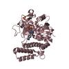
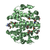
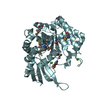
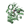
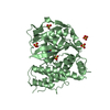
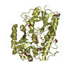
| Title | Klebsiella oxytoca NpsA N-terminal subdomain in space group C2 | ||||||
 Components Components | NpsA Adenylation Domain | ||||||
 Keywords Keywords |  BIOSYNTHETIC PROTEIN / adenylation / tilivalline / tilimycin / NRPS / nonribosomal peptide synthetase BIOSYNTHETIC PROTEIN / adenylation / tilivalline / tilimycin / NRPS / nonribosomal peptide synthetase | ||||||
| Function / homology |  Function and homology information Function and homology information | ||||||
| Biological species |  Klebsiella oxytoca (bacteria) Klebsiella oxytoca (bacteria) | ||||||
| Method | X-RAY DIFFRACTION / SYNCHROTRON / MOLECULAR REPLACEMENT / Resolution: 1.84 Å | ||||||
 Authors Authors | Kreitler, D.F. / Gulick, A.M. | ||||||
| Funding support |  United States, 1items United States, 1items
| ||||||
 Citation Citation | Journal: Acs Infect Dis. / Year: 2020 Title: Biosynthesis, Mechanism of Action, and Inhibition of the Enterotoxin Tilimycin Produced by the Opportunistic PathogenKlebsiella oxytoca. Authors: Alexander, E.M. / Kreitler, D.F. / Guidolin, V. / Hurben, A.K. / Drake, E. / Villalta, P.W. / Balbo, S. / Gulick, A.M. / Aldrich, C.C. | ||||||
| History |
|
- Structure visualization
| Structure viewer | Molecule: MolmilJmol/JSmol |
|---|
- Downloads & links
-Download
| PDBx/mmCIF format | 6vht.cif.gz | 175.6 KB | Display | PDBx/mmCIF format |
|---|---|---|---|---|
| PDB format | pdb6vht.ent.gz | 122.2 KB | Display | PDB format |
| PDBx/mmJSON format | 6vht.json.gz | Tree view | PDBx/mmJSON format | |
| Others |  Other downloads Other downloads |
-Validation report
| Arichive directory | https://data.pdbj.org/pub/pdb/validation_reports/vh/6vhtftp://data.pdbj.org/pub/pdb/validation_reports/vh/6vht | HTTPS FTP |
|---|
-Related structure data
| Related structure data | 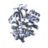 6vhuC  6vhvC 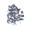 6vhwC  6vhxC 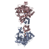 6vhyC 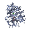 6vhzC 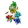 2vsqS S: Starting model for refinement C: citing same article ( |
|---|---|
| Similar structure data |


-Links
 PDBj
PDBj


- Assembly
| Deposited unit | 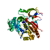
| ||||||||||||
|---|---|---|---|---|---|---|---|---|---|---|---|---|---|
| 1 |
| ||||||||||||
| 2 | 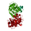
| ||||||||||||
| Unit cell |
|
-Components
| #1: Protein | Mass: 43960.355 Da / Num. of mol.: 1 / Mutation: E312A, E313A, Q314A Source method: isolated from a genetically manipulated source Source: (gene. exp.) Klebsiella oxytoca (bacteria) / Gene: NPSA / Plasmid: pET15 / Details (production host): N-term TEV tag / Production host: Escherichia coli (E. coli) / References: UniProt: A0A2U4DY99 | ||||
|---|---|---|---|---|---|
| #2: Chemical | ChemComp-BR / Bromide  Mass: 79.904 Da / Num. of mol.: 1 / Source method: obtained synthetically / Formula: Br Mass: 79.904 Da / Num. of mol.: 1 / Source method: obtained synthetically / Formula: Br | ||||
| #3: Chemical | Chloride  Mass: 35.453 Da / Num. of mol.: 2 / Source method: obtained synthetically / Formula: Cl Mass: 35.453 Da / Num. of mol.: 2 / Source method: obtained synthetically / Formula: Cl#4: Water | ChemComp-HOH / | Water Mass: 18.015 Da / Num. of mol.: 128 / Source method: isolated from a natural source / Formula: H2O Mass: 18.015 Da / Num. of mol.: 128 / Source method: isolated from a natural source / Formula: H2OHas ligand of interest | N | |
-Experimental details
-Experiment
| Experiment | Method: X-RAY DIFFRACTION / Number of used crystals: 1 |
|---|
- Sample preparation
| Crystal | Density Matthews: 2.06 Å3/Da / Density % sol: 40.16 % / Description: 3D |
|---|---|
| Crystal grow | Temperature: 293 K / Method: vapor diffusion, hanging drop Details: well solution: 100 mM HEPES pH 7.5, 150 mM KBr, 30% w/v PEG3350 protein solution (15 mg/mL): 50 mM HEPES pH 8.0, 150 mM NaCl, 0.2 mM TCEP hanging drops: 1 uL protein solution, 1 uL well solution PH range: 7.5-8 |
-Data collection
| Diffraction | Mean temperature: 100 K / Serial crystal experiment: N |
|---|---|
| Diffraction source | Source: SYNCHROTRON / Site: APS / Beamline: 23-ID-D / Wavelength: 1.03322 Å |
| Detector | Type: DECTRIS PILATUS3 6M / Detector: PIXEL / Date: Mar 25, 2019 |
| Radiation | Protocol: SINGLE WAVELENGTH / Monochromatic (M) / Laue (L): M / Scattering type: x-ray |
| Radiation wavelength | Wavelength: 1.03322 Å / Relative weight: 1 |
| Reflection | Resolution: 1.84→46.76 Å / Num. obs: 29872 / % possible obs: 96.3 % / Redundancy: 6.8 % / Biso Wilson estimate: 36.28 Å2 / CC1/2: 0.999 / Rpim(I) all: 0.034 / Rrim(I) all: 0.09 / Rsym value: 0.083 / Net I/σ(I): 10.2 |
| Reflection shell | Resolution: 1.84→1.87 Å / Redundancy: 6.5 % / Mean I/σ(I) obs: 0.9 / Num. unique obs: 1552 / CC1/2: 0.335 / Rpim(I) all: 0.886 / Rrim(I) all: 2.286 / Rsym value: 2.103 / % possible all: 96.9 |
- Processing
| Software |
| ||||||||||||||||||||||||||||||||||||||||||||||||||||||||||||||||||||||||||||||||||||
|---|---|---|---|---|---|---|---|---|---|---|---|---|---|---|---|---|---|---|---|---|---|---|---|---|---|---|---|---|---|---|---|---|---|---|---|---|---|---|---|---|---|---|---|---|---|---|---|---|---|---|---|---|---|---|---|---|---|---|---|---|---|---|---|---|---|---|---|---|---|---|---|---|---|---|---|---|---|---|---|---|---|---|---|---|---|
| Refinement | Method to determine structure: MOLECULAR REPLACEMENT Starting model: 2vsq Resolution: 1.84→46.76 Å / SU ML: 0.2829 / Cross valid method: FREE R-VALUE / σ(F): 1.34 / Phase error: 30.2238
| ||||||||||||||||||||||||||||||||||||||||||||||||||||||||||||||||||||||||||||||||||||
| Solvent computation | Shrinkage radii: 0.9 Å / VDW probe radii: 1.11 Å | ||||||||||||||||||||||||||||||||||||||||||||||||||||||||||||||||||||||||||||||||||||
| Displacement parameters | Biso mean: 52.98 Å2 | ||||||||||||||||||||||||||||||||||||||||||||||||||||||||||||||||||||||||||||||||||||
| Refinement step | Cycle: LAST / Resolution: 1.84→46.76 Å
| ||||||||||||||||||||||||||||||||||||||||||||||||||||||||||||||||||||||||||||||||||||
| Refine LS restraints |
| ||||||||||||||||||||||||||||||||||||||||||||||||||||||||||||||||||||||||||||||||||||
| LS refinement shell |
| ||||||||||||||||||||||||||||||||||||||||||||||||||||||||||||||||||||||||||||||||||||
| Refinement TLS params. | Method: refined / Refine-ID: X-RAY DIFFRACTION
| ||||||||||||||||||||||||||||||||||||||||||||||||||||||||||||||||||||||||||||||||||||
| Refinement TLS group |
|
