 Movie
Movie Controller


+ Open data

- Basic information
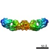
| Entry | Database: PDB / ID: 6vfr | ||||||||||||
|---|---|---|---|---|---|---|---|---|---|---|---|---|---|
| Title | Crystal structure of human protocadherin 18 EC1-EC4 | ||||||||||||
 Components Components | Protocadherin-18 | ||||||||||||
 Keywords Keywords | CELL ADHESION / cadherin extracellular region / non-clustered delta2 family protocadherin / homophilic adhesion/recognition calcium-dependent adhesion molecule | ||||||||||||
| Function / homology |  Function and homology information Function and homology informationhomophilic cell adhesion via plasma membrane adhesion molecules / plasma membrane => GO:0005886 / brain development / cell adhesion / calcium ion bindingSimilarity search - Function | ||||||||||||
| Biological species |  Homo sapiens (human) Homo sapiens (human) | ||||||||||||
| Method | X-RAY DIFFRACTION / SYNCHROTRON / MOLECULAR REPLACEMENT / Resolution: 2.79 Å | ||||||||||||
 Authors Authors | Harrison, O.J. / Brasch, J. / Shapiro, L. | ||||||||||||
| Funding support |  United States, 3items United States, 3items
| ||||||||||||
 Citation Citation | Journal: Cell Rep / Year: 2020 Title: Family-wide Structural and Biophysical Analysis of Binding Interactions among Non-clustered δ-Protocadherins. Authors: Oliver J Harrison / Julia Brasch / Phinikoula S Katsamba / Goran Ahlsen / Alex J Noble / Hanbin Dan / Rosemary V Sampogna / Clinton S Potter / Bridget Carragher / Barry Honig / Lawrence Shapiro / Abstract: Non-clustered δ1- and δ2-protocadherins, close relatives of clustered protocadherins, function in cell adhesion and motility and play essential roles in neural patterning. To understand the ...Non-clustered δ1- and δ2-protocadherins, close relatives of clustered protocadherins, function in cell adhesion and motility and play essential roles in neural patterning. To understand the molecular interactions underlying these functions, we used solution biophysics to characterize binding of δ1- and δ2-protocadherins, determined crystal structures of ectodomain complexes from each family, and assessed ectodomain assembly in reconstituted intermembrane junctions by cryoelectron tomography (cryo-ET). Homophilic trans (cell-cell) interactions were preferred for all δ-protocadherins, with additional weaker heterophilic interactions observed exclusively within each subfamily. As expected, δ1- and δ2-protocadherin trans dimers formed through antiparallel EC1-EC4 interfaces, like clustered protocadherins. However, no ectodomain-mediated cis (same-cell) interactions were detectable in solution; consistent with this, cryo-ET of reconstituted junctions revealed dense assemblies lacking the characteristic order observed for clustered protocadherins. Our results define non-clustered protocadherin binding properties and their structural basis, providing a foundation for interpreting their functional roles in neural patterning. | ||||||||||||
| History |
|
- Structure visualization
| Structure viewer | Molecule: MolmilJmol/JSmol |
|---|
- Downloads & links
-Download
| PDBx/mmCIF format | 6vfr.cif.gz | 411.7 KB | Display | PDBx/mmCIF format |
|---|---|---|---|---|
| PDB format | pdb6vfr.ent.gz | 275.6 KB | Display | PDB format |
| PDBx/mmJSON format | 6vfr.json.gz | Tree view | PDBx/mmJSON format | |
| Others |  Other downloads Other downloads |
-Validation report
| Arichive directory | https://data.pdbj.org/pub/pdb/validation_reports/vf/6vfrftp://data.pdbj.org/pub/pdb/validation_reports/vf/6vfr | HTTPS FTP |
|---|
-Related structure data
| Related structure data |  6vfpC  6vfqSC  6vftC  6vfuC  6vfvC  6vfwC  6vg1C  6vg4C S: Starting model for refinement C: citing same article ( |
|---|---|
| Similar structure data |


-Links
 PDBj
PDBj
- Assembly
| Deposited unit | 
| |||||||||||||||||||||||||||||||||||||||||||||||||||||||||||||||||||||||||||||||||||||||||||||||||||||||
|---|---|---|---|---|---|---|---|---|---|---|---|---|---|---|---|---|---|---|---|---|---|---|---|---|---|---|---|---|---|---|---|---|---|---|---|---|---|---|---|---|---|---|---|---|---|---|---|---|---|---|---|---|---|---|---|---|---|---|---|---|---|---|---|---|---|---|---|---|---|---|---|---|---|---|---|---|---|---|---|---|---|---|---|---|---|---|---|---|---|---|---|---|---|---|---|---|---|---|---|---|---|---|---|---|
| 1 |
| |||||||||||||||||||||||||||||||||||||||||||||||||||||||||||||||||||||||||||||||||||||||||||||||||||||||
| Unit cell |
| |||||||||||||||||||||||||||||||||||||||||||||||||||||||||||||||||||||||||||||||||||||||||||||||||||||||
| Noncrystallographic symmetry (NCS) | NCS domain:
NCS domain segments:
|