 Movie
Movie Controller

[English] 日本語
 Yorodumi
- PDB-6vea: Structure of the Glutamate-Like Receptor GLR3.2 ligand-binding do... -
+ Open data

- Basic information
| Entry | Database: PDB / ID: 6vea | |||||||||||||||
|---|---|---|---|---|---|---|---|---|---|---|---|---|---|---|---|---|
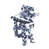
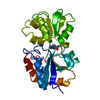
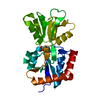
| Title | Structure of the Glutamate-Like Receptor GLR3.2 ligand-binding domain in complex with Glycine | |||||||||||||||
 Components Components | Glutamate receptor 3.2 | |||||||||||||||
 Keywords Keywords |  MEMBRANE PROTEIN / ligand-binding domain / Glutamate like receptor / Ion channel / Arabidopsis MEMBRANE PROTEIN / ligand-binding domain / Glutamate like receptor / Ion channel / Arabidopsis | |||||||||||||||
| Function / homology |  Function and homology informationglutamate receptor activity / ligand-gated monoatomic ion channel activity / calcium-mediated signaling / cellular response to amino acid stimulus / calcium channel activity / calcium ion transport / signaling receptor activity / plasma membrane Function and homology informationglutamate receptor activity / ligand-gated monoatomic ion channel activity / calcium-mediated signaling / cellular response to amino acid stimulus / calcium channel activity / calcium ion transport / signaling receptor activity / plasma membraneSimilarity search - Function | |||||||||||||||
| Biological species |  Arabidopsis thaliana (thale cress) Arabidopsis thaliana (thale cress) | |||||||||||||||
| Method | X-RAY DIFFRACTION / SYNCHROTRON / MOLECULAR REPLACEMENT / molecular replacement / Resolution: 1.58 Å | |||||||||||||||
 Authors Authors | Gangwar, S.P. / Green, M.N. / Yoder, J.B. / Sobolevsky, A.I. | |||||||||||||||
| Funding support |  United States, 4items United States, 4items
| |||||||||||||||
 Citation Citation | Journal: Structure / Year: 2021 Title: Structure of the Arabidopsis Glutamate Receptor-like Channel GLR3.2 Ligand-Binding Domain. Authors: Gangwar, S.P. / Green, M.N. / Michard, E. / Simon, A.A. / Feijo, J.A. / Sobolevsky, A.I. | |||||||||||||||
| History |
|
- Structure visualization
| Structure viewer | Molecule: MolmilJmol/JSmol |
|---|
- Downloads & links
-Download
| PDBx/mmCIF format | 6vea.cif.gz | 68.8 KB | Display | PDBx/mmCIF format |
|---|---|---|---|---|
| PDB format | pdb6vea.ent.gz | 46.5 KB | Display | PDB format |
| PDBx/mmJSON format | 6vea.json.gz | Tree view | PDBx/mmJSON format | |
| Others |  Other downloads Other downloads |
-Validation report
| Arichive directory | https://data.pdbj.org/pub/pdb/validation_reports/ve/6veaftp://data.pdbj.org/pub/pdb/validation_reports/ve/6vea | HTTPS FTP |
|---|
-Related structure data
| Related structure data |  6ve8SC S: Starting model for refinement C: citing same article ( |
|---|---|
| Similar structure data |


-Links
 PDBj
PDBj


- Assembly
| Deposited unit | 
| ||||||||
|---|---|---|---|---|---|---|---|---|---|
| 1 |
| ||||||||
| Unit cell |
|
-Components
| #1: Protein | Mass: 33778.094 Da / Num. of mol.: 1 / Fragment: ligand-binding domain Source method: isolated from a genetically manipulated source Source: (gene. exp.) Arabidopsis thaliana (thale cress) / Gene: GLR3.2, GLUR2, At4g35290, F23E12.150 / Plasmid: pET22b / Production host:  Escherichia coli (E. coli) / Strain (production host): Origami B (DE3) / References: UniProt: Q93YT1 Escherichia coli (E. coli) / Strain (production host): Origami B (DE3) / References: UniProt: Q93YT1 | ||||
|---|---|---|---|---|---|
| #2: Chemical | ChemComp-GLY / Glycine  Type: peptide linking / Mass: 75.067 Da / Num. of mol.: 1 / Source method: isolated from a natural source / Formula: C2H5NO2 / Feature type: SUBJECT OF INVESTIGATION Type: peptide linking / Mass: 75.067 Da / Num. of mol.: 1 / Source method: isolated from a natural source / Formula: C2H5NO2 / Feature type: SUBJECT OF INVESTIGATION | ||||
| #3: Chemical | ChemComp-NA /   Mass: 22.990 Da / Num. of mol.: 1 / Source method: isolated from a natural source / Formula: Na Mass: 22.990 Da / Num. of mol.: 1 / Source method: isolated from a natural source / Formula: Na | ||||
| #4: Chemical | 2-Mercaptoethanol  Mass: 78.133 Da / Num. of mol.: 2 / Source method: obtained synthetically / Formula: C2H6OS Mass: 78.133 Da / Num. of mol.: 2 / Source method: obtained synthetically / Formula: C2H6OS#5: Water | ChemComp-HOH / | Water Mass: 18.015 Da / Num. of mol.: 191 / Source method: isolated from a natural source / Formula: H2O Mass: 18.015 Da / Num. of mol.: 191 / Source method: isolated from a natural source / Formula: H2OHas ligand of interest | Y | |
-Experimental details
-Experiment
| Experiment | Method: X-RAY DIFFRACTION / Number of used crystals: 1 |
|---|
- Sample preparation
| Crystal | Density Matthews: 2.15 Å3/Da / Density % sol: 42.86 % |
|---|---|
| Crystal grow | Temperature: 277 K / Method: vapor diffusion, hanging drop / pH: 4.6 Details: 22 % PEG 4000, 0.1 M Ammonium acetate, 0.1 M Sodium acetate pH 4.6 |
-Data collection
| Diffraction | Mean temperature: 100 K / Serial crystal experiment: N | |||||||||||||||||||||||||||
|---|---|---|---|---|---|---|---|---|---|---|---|---|---|---|---|---|---|---|---|---|---|---|---|---|---|---|---|---|
| Diffraction source | Source: SYNCHROTRON / Site: APS / Beamline: 24-ID-C / Wavelength: 0.9791 Å | |||||||||||||||||||||||||||
| Detector | Type: DECTRIS PILATUS 6M / Detector: PIXEL / Date: Nov 6, 2019 | |||||||||||||||||||||||||||
| Radiation | Protocol: SINGLE WAVELENGTH / Monochromatic (M) / Laue (L): M / Scattering type: x-ray | |||||||||||||||||||||||||||
| Radiation wavelength | Wavelength: 0.9791 Å / Relative weight: 1 | |||||||||||||||||||||||||||
| Reflection | Resolution: 1.58→47.39 Å / Num. obs: 32133 / % possible obs: 99.2 % / Redundancy: 4.6 % / CC1/2: 0.999 / Rmerge(I) obs: 0.062 / Rpim(I) all: 0.031 / Rrim(I) all: 0.069 / Net I/σ(I): 14.9 / Num. measured all: 146995 / Scaling rejects: 8 | |||||||||||||||||||||||||||
| Reflection shell | Diffraction-ID: 1
|
-Phasing
| Phasing | Method: molecular replacement |
|---|
- Processing
| Software |
| ||||||||||||||||||||||||||||||||||||||||||||||||||||||||||||
|---|---|---|---|---|---|---|---|---|---|---|---|---|---|---|---|---|---|---|---|---|---|---|---|---|---|---|---|---|---|---|---|---|---|---|---|---|---|---|---|---|---|---|---|---|---|---|---|---|---|---|---|---|---|---|---|---|---|---|---|---|---|
| Refinement | Method to determine structure: MOLECULAR REPLACEMENT Starting model: 6VE8 Resolution: 1.58→40.23 Å / Cor.coef. Fo:Fc: 0.971 / Cor.coef. Fo:Fc free: 0.964 / SU B: 1.601 / SU ML: 0.055 / Cross valid method: THROUGHOUT / σ(F): 0 / ESU R: 0.077 / ESU R Free: 0.077 / Stereochemistry target values: MAXIMUM LIKELIHOOD Details: HYDROGENS HAVE BEEN ADDED IN THE RIDING POSITIONS U VALUES : REFINED INDIVIDUALLY
| ||||||||||||||||||||||||||||||||||||||||||||||||||||||||||||
| Solvent computation | Ion probe radii: 0.8 Å / Shrinkage radii: 0.8 Å / VDW probe radii: 1.2 Å / Solvent model: MASK | ||||||||||||||||||||||||||||||||||||||||||||||||||||||||||||
| Displacement parameters | Biso max: 71.87 Å2 / Biso mean: 19.238 Å2 / Biso min: 10.61 Å2
| ||||||||||||||||||||||||||||||||||||||||||||||||||||||||||||
| Refinement step | Cycle: final / Resolution: 1.58→40.23 Å
| ||||||||||||||||||||||||||||||||||||||||||||||||||||||||||||
| Refine LS restraints |
| ||||||||||||||||||||||||||||||||||||||||||||||||||||||||||||
| LS refinement shell | Resolution: 1.58→1.621 Å / Rfactor Rfree error: 0 / Total num. of bins used: 20
|
