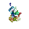
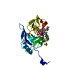
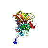
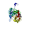
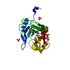
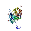
Entry Database : PDB / ID : 6vdoTitle HCV NS3/4A protease A156T, D168E double mutant NS3 protease Keywords / / / / Function / homology Function Domain/homology Component
/ / / / / / / / / / / / / / / / / / / / / / / / / / / / / / / / / / / / / / / / / / / / / / / / / / / / / / / / / / / / / / / / / / / / / / / / / / / / / / / / / / / / / / / / / / / / / / / / / / / / / / / / / / / / / / / Biological species Method / / Resolution : 2.11 Å Authors Timm, J. / Schiffer, C.A. Funding support Organization Grant number Country National Institutes of Health/National Institute Of Allergy and Infectious Diseases (NIH/NIAID) R01AI085051
Journal : To Be Published Title : apo structure of HCV NS3/4A protease A156T;D168E double mutantAuthors : Timm, J. / Zephyr, J. / Hou, S. / Kurt Yilmaz, N. / Schiffer, C.A. History Deposition Dec 27, 2019 Deposition site / Processing site Revision 1.0 Dec 30, 2020 Provider / Type Revision 1.1 Oct 11, 2023 Group / Database references / Refinement descriptionCategory chem_comp_atom / chem_comp_bond ... chem_comp_atom / chem_comp_bond / database_2 / pdbx_initial_refinement_model Item / _database_2.pdbx_database_accession
Show all Show less
 Movie
Movie Controller


 Open data
 Basic information
 Components
 Keywords
 VIRAL PROTEIN / HCV protease / NS34A protease / A156T / D168E
 Function and homology information
 Hepacivirus C
 Authors
 United States, 1items
  Citation
 Structure visualization
 Downloads & links
 Other downloads
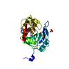


 PDBj
PDBj


 Assembly


 Mass: 65.409 Da / Num. of mol.: 1 / Source method: obtained synthetically / Formula: Zn
 Mass: 78.133 Da / Num. of mol.: 1 / Source method: obtained synthetically / Formula: C2H6OS
 Mass: 96.063 Da / Num. of mol.: 3 / Source method: obtained synthetically / Formula: SO4
 Mass: 18.015 Da / Num. of mol.: 141 / Source method: isolated from a natural source / Formula: H2O
 Sample preparation
 Processing