 Movie
Movie Controller


+ Open data

- Basic information
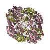
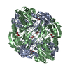
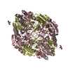
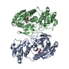
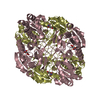
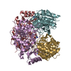
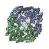
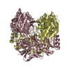
| Entry | Database: PDB / ID: 6v5f | ||||||
|---|---|---|---|---|---|---|---|
| Title | L-asparaginase from Escherichia coli | ||||||
 Components Components | L-asparaginase 2 Asparaginase Asparaginase | ||||||
 Keywords Keywords | HYDROLASE / L-asparaginase / catalytic mechanism / leukemia | ||||||
| Function / homology |  Function and homology information Function and homology informationasparagine catabolic process / asparaginase / asparaginase activity / outer membrane-bounded periplasmic space / protein homotetramerization / periplasmic space / protein-containing complex / identical protein bindingSimilarity search - Function | ||||||
| Biological species |  Escherichia coli (E. coli) Escherichia coli (E. coli) | ||||||
| Method | X-RAY DIFFRACTION / SYNCHROTRON / MOLECULAR REPLACEMENT / molecular replacement / Resolution: 2.1 Å | ||||||
 Authors Authors | Souza, T.A.C.B. / Morais, S.B. / Morini, F.S. / Meissner, G.O. | ||||||
 Citation Citation | Journal: To Be Published Title: L-asparaginase from Escherichia coli: Novel insights about the behavior of flexible part of the active site Authors: Souza, T.A.C.B. / Morais, S.B. / Morini, F.S. / Meissner, G.O. | ||||||
| History |
|
- Structure visualization
| Structure viewer | Molecule: MolmilJmol/JSmol |
|---|
- Downloads & links
-Download
| PDBx/mmCIF format | 6v5f.cif.gz | 239.6 KB | Display | PDBx/mmCIF format |
|---|---|---|---|---|
| PDB format | pdb6v5f.ent.gz | 193 KB | Display | PDB format |
| PDBx/mmJSON format | 6v5f.json.gz | Tree view | PDBx/mmJSON format | |
| Others |  Other downloads Other downloads |
-Validation report
| Arichive directory | https://data.pdbj.org/pub/pdb/validation_reports/v5/6v5fftp://data.pdbj.org/pub/pdb/validation_reports/v5/6v5f | HTTPS FTP |
|---|
-Related structure data
| Related structure data |  7m11C  3ecaS S: Starting model for refinement C: citing same article ( |
|---|---|
| Similar structure data |

-Links
 PDBj
PDBj
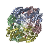
- Assembly
| Deposited unit | 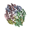
| |||||||||||||||||||||||||||||||||||||||||||||||||||||||||||||||||||||||||||||||
|---|---|---|---|---|---|---|---|---|---|---|---|---|---|---|---|---|---|---|---|---|---|---|---|---|---|---|---|---|---|---|---|---|---|---|---|---|---|---|---|---|---|---|---|---|---|---|---|---|---|---|---|---|---|---|---|---|---|---|---|---|---|---|---|---|---|---|---|---|---|---|---|---|---|---|---|---|---|---|---|---|
| 1 |
| |||||||||||||||||||||||||||||||||||||||||||||||||||||||||||||||||||||||||||||||
| Unit cell |
| |||||||||||||||||||||||||||||||||||||||||||||||||||||||||||||||||||||||||||||||
| Noncrystallographic symmetry (NCS) | NCS domain:
NCS domain segments:
|