 Movie
Movie Controller

[English] 日本語
 Yorodumi
- PDB-6v4w: The crystal structure of a beta-lactamase from Chitinophaga pinen... -
+ Open data

- Basic information
| Entry | Database: PDB / ID: 6v4w | ||||||
|---|---|---|---|---|---|---|---|
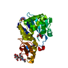
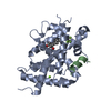
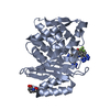
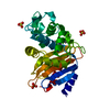
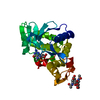
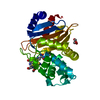
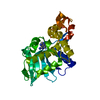
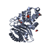
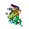
| Title | The crystal structure of a beta-lactamase from Chitinophaga pinensis DSM 2588 | ||||||
 Components Components | Beta-lactamase | ||||||
 Keywords Keywords | HYDROLASE / Class A beta-lactamase / Structural Genomics / Center for Structural Genomics of Infectious Diseases / CSGID | ||||||
| Function / homology |  Function and homology information Function and homology informationbeta-lactam antibiotic catabolic process / beta-lactamase activity / beta-lactamase / response to antibioticSimilarity search - Function | ||||||
| Biological species |  Chitinophaga pinensis (bacteria) Chitinophaga pinensis (bacteria) | ||||||
| Method | X-RAY DIFFRACTION / SYNCHROTRON / MOLECULAR REPLACEMENT / Resolution: 1.29 Å | ||||||
 Authors Authors | Tan, K. / Welk, L. / Endres, M. / Joachimiak, A. / Center for Structural Genomics of Infectious Diseases (CSGID) | ||||||
| Funding support |  United States, 1items United States, 1items
| ||||||
 Citation Citation | Journal: To Be Published Title: The crystal structure of a beta-lactamase from Chitinophaga pinensis DSM 2588 Authors: Tan, K. / Welk, L. / Endres, M. / Joachimiak, A. | ||||||
| History |
|
- Structure visualization
| Structure viewer | Molecule: MolmilJmol/JSmol |
|---|
- Downloads & links
-Download
| PDBx/mmCIF format | 6v4w.cif.gz | 280.5 KB | Display | PDBx/mmCIF format |
|---|---|---|---|---|
| PDB format | pdb6v4w.ent.gz | 187.1 KB | Display | PDB format |
| PDBx/mmJSON format | 6v4w.json.gz | Tree view | PDBx/mmJSON format | |
| Others |  Other downloads Other downloads |
-Validation report
| Arichive directory | https://data.pdbj.org/pub/pdb/validation_reports/v4/6v4wftp://data.pdbj.org/pub/pdb/validation_reports/v4/6v4w | HTTPS FTP |
|---|
-Related structure data
| Related structure data |  5g58S S: Starting model for refinement |
|---|---|
| Similar structure data | |
| Other databases |

-Links
 PDBj
PDBj


- Assembly
| Deposited unit | 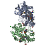
| ||||||||||||
|---|---|---|---|---|---|---|---|---|---|---|---|---|---|
| 1 | 
| ||||||||||||
| 2 | 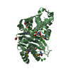
| ||||||||||||
| Unit cell |
|
-Components
-Protein , 1 types, 2 molecules AB
| #1: Protein | Mass: 30552.516 Da / Num. of mol.: 2 Source method: isolated from a genetically manipulated source Source: (gene. exp.) Chitinophaga pinensis (strain ATCC 43595 / DSM 2588 / NCIB 11800 / UQM 2034) (bacteria)Strain: ATCC 43595 / DSM 2588 / NCIB 11800 / UQM 2034 / Gene: Cpin_1435 / Plasmid: pMCSG53 / Production host: Escherichia coli (E. coli) / Strain (production host): BL21-Gold(DE3) / References: UniProt: C7PRN9, beta-lactamase |
|---|
-Non-polymers , 5 types, 552 molecules 


| #2: Chemical | Acetate Mass: 59.044 Da / Num. of mol.: 3 / Source method: obtained synthetically / Formula: C2H3O2 Mass: 59.044 Da / Num. of mol.: 3 / Source method: obtained synthetically / Formula: C2H3O2#3: Chemical | ChemComp-CL / Chloride Mass: 35.453 Da / Num. of mol.: 8 / Source method: obtained synthetically / Formula: Cl Mass: 35.453 Da / Num. of mol.: 8 / Source method: obtained synthetically / Formula: Cl#4: Chemical | ChemComp-MES / | MES (buffer) Mass: 195.237 Da / Num. of mol.: 1 / Source method: obtained synthetically / Formula: C6H13NO4S / Comment: pH buffer*YM Mass: 195.237 Da / Num. of mol.: 1 / Source method: obtained synthetically / Formula: C6H13NO4S / Comment: pH buffer*YM#5: Chemical | ChemComp-FMT / | Formic acid Mass: 46.025 Da / Num. of mol.: 1 / Source method: obtained synthetically / Formula: CH2O2 Mass: 46.025 Da / Num. of mol.: 1 / Source method: obtained synthetically / Formula: CH2O2#6: Water | ChemComp-HOH / | WaterMass: 18.015 Da / Num. of mol.: 539 / Source method: isolated from a natural source / Formula: H2O |
|---|
-Details
| Has ligand of interest | N |
|---|
-Experimental details
-Experiment
| Experiment | Method: X-RAY DIFFRACTION / Number of used crystals: 1 |
|---|
- Sample preparation
| Crystal | Density Matthews: 2.1 Å3/Da / Density % sol: 41.38 % |
|---|---|
| Crystal grow | Temperature: 296 K / Method: vapor diffusion, sitting drop / pH: 6.5 Details: 0.2 M Magnesium Chloride, 0.1 M MES:NaOH, 25% (w/v) PEG 4000 |
-Data collection
| Diffraction | Mean temperature: 100 K / Serial crystal experiment: N |
|---|---|
| Diffraction source | Source: SYNCHROTRON / Site: APS / Beamline: 19-ID / Wavelength: 0.97918 Å |
| Detector | Type: DECTRIS PILATUS3 S 6M / Detector: PIXEL / Date: Nov 7, 2018 |
| Radiation | Monochromator: Si 111 / Protocol: SINGLE WAVELENGTH / Monochromatic (M) / Laue (L): M / Scattering type: x-ray |
| Radiation wavelength | Wavelength: 0.97918 Å / Relative weight: 1 |
| Reflection | Resolution: 1.28→50 Å / Num. obs: 127393 / % possible obs: 97.7 % / Observed criterion σ(I): -3 / Redundancy: 3 % / Biso Wilson estimate: 15.83 Å2 / Rmerge(I) obs: 0.099 / Rpim(I) all: 0.067 / Rrim(I) all: 0.121 / Χ2: 3.11 / Net I/σ(I): 23.1 |
| Reflection shell | Resolution: 1.28→1.3 Å / Redundancy: 2.5 % / Rmerge(I) obs: 0.771 / Mean I/σ(I) obs: 1.1 / Num. unique obs: 6253 / CC1/2: 0.457 / Rpim(I) all: 0.563 / Rrim(I) all: 0.961 / Χ2: 3.1 / % possible all: 97.2 |
- Processing
| Software |
| |||||||||||||||||||||||||||||||||||||||||||||||||||||||||||||||||||||||||||||||||||||||||||||||||||||||||||||||||||||||||||||||||||||||||||||||||||||||||||||||||||||||||||||||||||||||||||||||||||||||||||||||||||||||||
|---|---|---|---|---|---|---|---|---|---|---|---|---|---|---|---|---|---|---|---|---|---|---|---|---|---|---|---|---|---|---|---|---|---|---|---|---|---|---|---|---|---|---|---|---|---|---|---|---|---|---|---|---|---|---|---|---|---|---|---|---|---|---|---|---|---|---|---|---|---|---|---|---|---|---|---|---|---|---|---|---|---|---|---|---|---|---|---|---|---|---|---|---|---|---|---|---|---|---|---|---|---|---|---|---|---|---|---|---|---|---|---|---|---|---|---|---|---|---|---|---|---|---|---|---|---|---|---|---|---|---|---|---|---|---|---|---|---|---|---|---|---|---|---|---|---|---|---|---|---|---|---|---|---|---|---|---|---|---|---|---|---|---|---|---|---|---|---|---|---|---|---|---|---|---|---|---|---|---|---|---|---|---|---|---|---|---|---|---|---|---|---|---|---|---|---|---|---|---|---|---|---|---|---|---|---|---|---|---|---|---|---|---|---|---|---|---|---|---|
| Refinement | Method to determine structure: MOLECULAR REPLACEMENT Starting model: 5G58 Resolution: 1.29→38.48 Å / SU ML: 0.1379 / Cross valid method: FREE R-VALUE / σ(F): 1.34 / Phase error: 18.6195
| |||||||||||||||||||||||||||||||||||||||||||||||||||||||||||||||||||||||||||||||||||||||||||||||||||||||||||||||||||||||||||||||||||||||||||||||||||||||||||||||||||||||||||||||||||||||||||||||||||||||||||||||||||||||||
| Solvent computation | Shrinkage radii: 0.9 Å / VDW probe radii: 1.11 Å | |||||||||||||||||||||||||||||||||||||||||||||||||||||||||||||||||||||||||||||||||||||||||||||||||||||||||||||||||||||||||||||||||||||||||||||||||||||||||||||||||||||||||||||||||||||||||||||||||||||||||||||||||||||||||
| Displacement parameters | Biso mean: 21.15 Å2 | |||||||||||||||||||||||||||||||||||||||||||||||||||||||||||||||||||||||||||||||||||||||||||||||||||||||||||||||||||||||||||||||||||||||||||||||||||||||||||||||||||||||||||||||||||||||||||||||||||||||||||||||||||||||||
| Refinement step | Cycle: LAST / Resolution: 1.29→38.48 Å
| |||||||||||||||||||||||||||||||||||||||||||||||||||||||||||||||||||||||||||||||||||||||||||||||||||||||||||||||||||||||||||||||||||||||||||||||||||||||||||||||||||||||||||||||||||||||||||||||||||||||||||||||||||||||||
| Refine LS restraints |
| |||||||||||||||||||||||||||||||||||||||||||||||||||||||||||||||||||||||||||||||||||||||||||||||||||||||||||||||||||||||||||||||||||||||||||||||||||||||||||||||||||||||||||||||||||||||||||||||||||||||||||||||||||||||||
| LS refinement shell |
|
