 Movie
Movie Controller

[English] 日本語
 Yorodumi
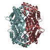
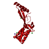
- PDB-6uxt: Crystal structure of unknown function protein yfdX from Shigella ... -
+ Open data

- Basic information
| Entry | Database: PDB / ID: 6uxt | ||||||
|---|---|---|---|---|---|---|---|
| Title | Crystal structure of unknown function protein yfdX from Shigella flexneri | ||||||
 Components Components | YfdX protein | ||||||
 Keywords Keywords |  STRUCTURAL GENOMICS / CSGID / Center for Structural Genomics of Infectious Diseases STRUCTURAL GENOMICS / CSGID / Center for Structural Genomics of Infectious Diseases | ||||||
| Function / homology |  Function and homology information Function and homology informationYfdX protein domain / Single alpha-helices involved in coiled-coils or other helix-helix interfaces - #2140 / Uncharacterised protein family YfdX / YfdX protein / Single alpha-helices involved in coiled-coils or other helix-helix interfaces / Four Helix Bundle (Hemerythrin (Met), subunit A) / Helix non-globular / Special / Up-down Bundle / Mainly Alpha Similarity search - Domain/homology | ||||||
| Biological species |  Shigella flexneri 2a str. 2457T (bacteria) Shigella flexneri 2a str. 2457T (bacteria) | ||||||
| Method | X-RAY DIFFRACTION / SYNCHROTRON / MOLECULAR REPLACEMENT / Resolution: 1.797 Å | ||||||
 Authors Authors | Chang, C. / Skarina, T. / Savchenko, A. / Joachimiak, A. / Center for Structural Genomics of Infectious Diseases (CSGID) | ||||||
 Citation Citation | Journal: To Be Published Title: Crystal structure of unknown function protein yfdX from Shigella flexneri Authors: Chang, C. / Skarina, T. / Savchenko, A. / Joachimiak, A. / Center for Structural Genomics of Infectious Diseases (CSGID) | ||||||
| History |
|
- Structure visualization
| Structure viewer | Molecule: MolmilJmol/JSmol |
|---|
- Downloads & links
-Download
| PDBx/mmCIF format | 6uxt.cif.gz | 280.5 KB | Display | PDBx/mmCIF format |
|---|---|---|---|---|
| PDB format | pdb6uxt.ent.gz | 225.7 KB | Display | PDB format |
| PDBx/mmJSON format | 6uxt.json.gz | Tree view | PDBx/mmJSON format | |
| Others |  Other downloads Other downloads |
-Validation report
| Arichive directory | https://data.pdbj.org/pub/pdb/validation_reports/ux/6uxtftp://data.pdbj.org/pub/pdb/validation_reports/ux/6uxt | HTTPS FTP |
|---|
-Related structure data
| Related structure data | 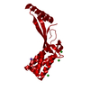 6a02S S: Starting model for refinement |
|---|---|
| Similar structure data | |
| Other databases |


-Links
 PDBj
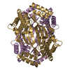
PDBj- Assembly
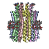
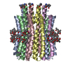
| Deposited unit | 
| ||||||||
|---|---|---|---|---|---|---|---|---|---|
| 1 | 
| ||||||||
| 2 | 
| ||||||||
| Unit cell |
|
-Components
| #1: Protein | Mass: 20297.846 Da / Num. of mol.: 8 Source method: isolated from a genetically manipulated source Source: (gene. exp.) Shigella flexneri 2a str. 2457T (bacteria)Gene: yfdX, / Production host: Escherichia coli BL21(DE3) (bacteria) / References: UniProt: A0A2Y2P124#2: Chemical | Polyethylene glycol  Mass: 150.173 Da / Num. of mol.: 2 / Source method: obtained synthetically / Formula: C6H14O4 Mass: 150.173 Da / Num. of mol.: 2 / Source method: obtained synthetically / Formula: C6H14O4#3: Chemical | ChemComp-PEG / | Diethylene glycol  Mass: 106.120 Da / Num. of mol.: 1 / Source method: obtained synthetically / Formula: C4H10O3 Mass: 106.120 Da / Num. of mol.: 1 / Source method: obtained synthetically / Formula: C4H10O3#4: Chemical | ChemComp-EPE / | HEPES  Mass: 238.305 Da / Num. of mol.: 1 / Source method: obtained synthetically / Formula: C8H18N2O4S / Comment: pH buffer*YM Mass: 238.305 Da / Num. of mol.: 1 / Source method: obtained synthetically / Formula: C8H18N2O4S / Comment: pH buffer*YM#5: Water | ChemComp-HOH / | Water Mass: 18.015 Da / Num. of mol.: 753 / Source method: isolated from a natural source / Formula: H2O Mass: 18.015 Da / Num. of mol.: 753 / Source method: isolated from a natural source / Formula: H2OHas ligand of interest | N | |
|---|
-Experimental details
-Experiment
| Experiment | Method: X-RAY DIFFRACTION / Number of used crystals: 1 |
|---|
- Sample preparation
| Crystal | Density Matthews: 2.69 Å3/Da / Density % sol: 54.24 % |
|---|---|
| Crystal grow | Temperature: 297 K / Method: vapor diffusion, hanging drop / pH: 6 / Details: 20% PEG3350, 0.2M ammonium citrate, 50mM MES |
-Data collection
| Diffraction | Mean temperature: 100 K / Serial crystal experiment: N | |||||||||||||||||||||||||||||||||||||||||||||||||||||||||||||||||||||||||||||||||||||||||||||||||||||||||||||||||||||||||||||||||||||||||||||||||||||||||||||||||||||||||||||||||||||||||||||
|---|---|---|---|---|---|---|---|---|---|---|---|---|---|---|---|---|---|---|---|---|---|---|---|---|---|---|---|---|---|---|---|---|---|---|---|---|---|---|---|---|---|---|---|---|---|---|---|---|---|---|---|---|---|---|---|---|---|---|---|---|---|---|---|---|---|---|---|---|---|---|---|---|---|---|---|---|---|---|---|---|---|---|---|---|---|---|---|---|---|---|---|---|---|---|---|---|---|---|---|---|---|---|---|---|---|---|---|---|---|---|---|---|---|---|---|---|---|---|---|---|---|---|---|---|---|---|---|---|---|---|---|---|---|---|---|---|---|---|---|---|---|---|---|---|---|---|---|---|---|---|---|---|---|---|---|---|---|---|---|---|---|---|---|---|---|---|---|---|---|---|---|---|---|---|---|---|---|---|---|---|---|---|---|---|---|---|---|---|---|---|
| Diffraction source | Source: SYNCHROTRON / Site: APS  / Beamline: 19-ID / Wavelength: 0.97918 Å / Beamline: 19-ID / Wavelength: 0.97918 Å | |||||||||||||||||||||||||||||||||||||||||||||||||||||||||||||||||||||||||||||||||||||||||||||||||||||||||||||||||||||||||||||||||||||||||||||||||||||||||||||||||||||||||||||||||||||||||||||
| Detector | Type: DECTRIS PILATUS3 X 6M / Detector: PIXEL / Date: Jun 11, 2019 | |||||||||||||||||||||||||||||||||||||||||||||||||||||||||||||||||||||||||||||||||||||||||||||||||||||||||||||||||||||||||||||||||||||||||||||||||||||||||||||||||||||||||||||||||||||||||||||
| Radiation | Protocol: SINGLE WAVELENGTH / Monochromatic (M) / Laue (L): M / Scattering type: x-ray | |||||||||||||||||||||||||||||||||||||||||||||||||||||||||||||||||||||||||||||||||||||||||||||||||||||||||||||||||||||||||||||||||||||||||||||||||||||||||||||||||||||||||||||||||||||||||||||
| Radiation wavelength | Wavelength: 0.97918 Å / Relative weight: 1 | |||||||||||||||||||||||||||||||||||||||||||||||||||||||||||||||||||||||||||||||||||||||||||||||||||||||||||||||||||||||||||||||||||||||||||||||||||||||||||||||||||||||||||||||||||||||||||||
| Reflection | Resolution: 1.797→50 Å / Num. obs: 143645 / % possible obs: 91.4 % / Redundancy: 3.7 % / Rmerge(I) obs: 0.062 / Rpim(I) all: 0.037 / Rrim(I) all: 0.073 / Χ2: 0.861 / Net I/σ(I): 7.1 | |||||||||||||||||||||||||||||||||||||||||||||||||||||||||||||||||||||||||||||||||||||||||||||||||||||||||||||||||||||||||||||||||||||||||||||||||||||||||||||||||||||||||||||||||||||||||||||
| Reflection shell | Diffraction-ID: 1
|
- Processing
| Software |
| ||||||||||||||||||||||||||||||||||||||||||||||||||||||||||||||||||||||||||||||||||||||||||||||||||||||||||||||||||||||||||||||||||||||||||||||||||||||||||||||||||||||||||||||||||||||||||
|---|---|---|---|---|---|---|---|---|---|---|---|---|---|---|---|---|---|---|---|---|---|---|---|---|---|---|---|---|---|---|---|---|---|---|---|---|---|---|---|---|---|---|---|---|---|---|---|---|---|---|---|---|---|---|---|---|---|---|---|---|---|---|---|---|---|---|---|---|---|---|---|---|---|---|---|---|---|---|---|---|---|---|---|---|---|---|---|---|---|---|---|---|---|---|---|---|---|---|---|---|---|---|---|---|---|---|---|---|---|---|---|---|---|---|---|---|---|---|---|---|---|---|---|---|---|---|---|---|---|---|---|---|---|---|---|---|---|---|---|---|---|---|---|---|---|---|---|---|---|---|---|---|---|---|---|---|---|---|---|---|---|---|---|---|---|---|---|---|---|---|---|---|---|---|---|---|---|---|---|---|---|---|---|---|---|---|---|
| Refinement | Method to determine structure: MOLECULAR REPLACEMENT Starting model: 6A02 Resolution: 1.797→43.517 Å / SU ML: 0.22 / Cross valid method: THROUGHOUT / σ(F): 1.98 / Phase error: 20.91
| ||||||||||||||||||||||||||||||||||||||||||||||||||||||||||||||||||||||||||||||||||||||||||||||||||||||||||||||||||||||||||||||||||||||||||||||||||||||||||||||||||||||||||||||||||||||||||
| Solvent computation | Shrinkage radii: 0.9 Å / VDW probe radii: 1.11 Å | ||||||||||||||||||||||||||||||||||||||||||||||||||||||||||||||||||||||||||||||||||||||||||||||||||||||||||||||||||||||||||||||||||||||||||||||||||||||||||||||||||||||||||||||||||||||||||
| Displacement parameters | Biso max: 100.5 Å2 / Biso mean: 32.3239 Å2 / Biso min: 15.35 Å2 | ||||||||||||||||||||||||||||||||||||||||||||||||||||||||||||||||||||||||||||||||||||||||||||||||||||||||||||||||||||||||||||||||||||||||||||||||||||||||||||||||||||||||||||||||||||||||||
| Refinement step | Cycle: final / Resolution: 1.797→43.517 Å
| ||||||||||||||||||||||||||||||||||||||||||||||||||||||||||||||||||||||||||||||||||||||||||||||||||||||||||||||||||||||||||||||||||||||||||||||||||||||||||||||||||||||||||||||||||||||||||
| LS refinement shell | Refine-ID: X-RAY DIFFRACTION / Rfactor Rfree error: 0
|
