 Movie
Movie Controller

[English] 日本語
 Yorodumi
- PDB-6us9: Influenza A M2 proton channel wild type TM domain bound to R-rima... -
+ Open data

- Basic information
| Entry | Database: PDB / ID: 6us9 | |||||||||
|---|---|---|---|---|---|---|---|---|---|---|
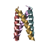
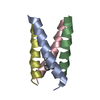
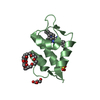
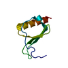
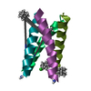
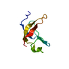
| Title | Influenza A M2 proton channel wild type TM domain bound to R-rimantadine | |||||||||
 Components Components | Matrix protein 2 | |||||||||
 Keywords Keywords |  MEMBRANE PROTEIN / proton channel / rimantadine MEMBRANE PROTEIN / proton channel / rimantadine | |||||||||
| Function / homology |  Function and homology information Function and homology informationproton transmembrane transporter activity / host cell membrane / : / protein complex oligomerization / monoatomic ion channel activity / virion membrane / membraneSimilarity search - Function | |||||||||
| Biological species |   Influenza A virus Influenza A virus | |||||||||
| Method | X-RAY DIFFRACTION / SYNCHROTRON / MOLECULAR REPLACEMENT / Resolution: 2 Å | |||||||||
 Authors Authors | Thomaston, J.L. / DeGrado, W.F. | |||||||||
| Funding support |  United States, 2items United States, 2items
| |||||||||
 Citation Citation | Journal: Biochemistry / Year: 2021 Title: Rimantadine Binds to and Inhibits the Influenza A M2 Proton Channel without Enantiomeric Specificity. Authors: Thomaston, J.L. / Samways, M.L. / Konstantinidi, A. / Ma, C. / Hu, Y. / Bruce Macdonald, H.E. / Wang, J. / Essex, J.W. / DeGrado, W.F. / Kolocouris, A. | |||||||||
| History |
|
- Structure visualization
| Structure viewer | Molecule: MolmilJmol/JSmol |
|---|
- Downloads & links
-Download
| PDBx/mmCIF format | 6us9.cif.gz | 94.3 KB | Display | PDBx/mmCIF format |
|---|---|---|---|---|
| PDB format | pdb6us9.ent.gz | 68.3 KB | Display | PDB format |
| PDBx/mmJSON format | 6us9.json.gz | Tree view | PDBx/mmJSON format | |
| Others |  Other downloads Other downloads |
-Validation report
| Arichive directory | https://data.pdbj.org/pub/pdb/validation_reports/us/6us9ftp://data.pdbj.org/pub/pdb/validation_reports/us/6us9 | HTTPS FTP |
|---|
-Related structure data
| Related structure data |  6us8C  6bklS S: Starting model for refinement C: citing same article ( |
|---|---|
| Similar structure data |


-Links
 PDBj
PDBj- Assembly
| Deposited unit | 
| ||||||||||||
|---|---|---|---|---|---|---|---|---|---|---|---|---|---|
| 1 | 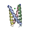
| ||||||||||||
| 2 | 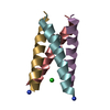
| ||||||||||||
| 3 | 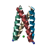
| ||||||||||||
| 4 | 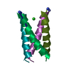
| ||||||||||||
| Unit cell |
|
-Components
| #1: Protein/peptide | Mass: 2754.340 Da / Num. of mol.: 16 / Source method: obtained synthetically Source: (synth.) Influenza A virus (A/Jinfang/132/2002(H3N2))References: UniProt: D5F6K1 #2: Chemical | ChemComp-RIM / Rimantadine  Mass: 179.302 Da / Num. of mol.: 4 / Source method: obtained synthetically / Formula: C12H21N / Feature type: SUBJECT OF INVESTIGATION / Comment: antivirus*YM Mass: 179.302 Da / Num. of mol.: 4 / Source method: obtained synthetically / Formula: C12H21N / Feature type: SUBJECT OF INVESTIGATION / Comment: antivirus*YM#3: Chemical | ChemComp-CL / Chloride  Mass: 35.453 Da / Num. of mol.: 4 / Source method: obtained synthetically / Formula: Cl Mass: 35.453 Da / Num. of mol.: 4 / Source method: obtained synthetically / Formula: Cl#4: Water | ChemComp-HOH / | Water Mass: 18.015 Da / Num. of mol.: 50 / Source method: isolated from a natural source / Formula: H2O Mass: 18.015 Da / Num. of mol.: 50 / Source method: isolated from a natural source / Formula: H2OHas ligand of interest | Y | |
|---|
-Experimental details
-Experiment
| Experiment | Method: X-RAY DIFFRACTION / Number of used crystals: 1 |
|---|
- Sample preparation
| Crystal | Density Matthews: 1.91 Å3/Da / Density % sol: 35.54 % |
|---|---|
| Crystal grow | Temperature: 293 K / Method: lipidic cubic phase / pH: 8.5 Details: monoolein, 0.015 M Tricine pH 8.5, 24% w/v PEG 4000, 50 mM MNG-3-C8, R-rimantadine |
-Data collection
| Diffraction | Mean temperature: 100 K / Serial crystal experiment: N |
|---|---|
| Diffraction source | Source: SYNCHROTRON / Site: ALS / Beamline: 8.3.1 / Wavelength: 1.1159 Å |
| Detector | Type: DECTRIS PILATUS3 S 6M / Detector: PIXEL / Date: Oct 20, 2018 |
| Radiation | Protocol: SINGLE WAVELENGTH / Monochromatic (M) / Laue (L): M / Scattering type: x-ray |
| Radiation wavelength | Wavelength: 1.1159 Å / Relative weight: 1 |
| Reflection | Resolution: 2→48.7 Å / Num. obs: 21661 / % possible obs: 96 % / Redundancy: 5.4 % / Biso Wilson estimate: 15.8024980126 Å2 / CC1/2: 0.997 / Rmerge(I) obs: 0.122 / Rpim(I) all: 0.056 / Net I/σ(I): 6.8 |
| Reflection shell | Resolution: 2→2.05 Å / Rmerge(I) obs: 0.494 / Mean I/σ(I) obs: 2.6 / Num. unique obs: 1558 / CC1/2: 0.95 / Rpim(I) all: 0.247 |
- Processing
| Software |
| ||||||||||||||||||||||||||||||||||||||||||||||||||||||||||||||||||||||||||||||||||||||||||||||||||
|---|---|---|---|---|---|---|---|---|---|---|---|---|---|---|---|---|---|---|---|---|---|---|---|---|---|---|---|---|---|---|---|---|---|---|---|---|---|---|---|---|---|---|---|---|---|---|---|---|---|---|---|---|---|---|---|---|---|---|---|---|---|---|---|---|---|---|---|---|---|---|---|---|---|---|---|---|---|---|---|---|---|---|---|---|---|---|---|---|---|---|---|---|---|---|---|---|---|---|---|
| Refinement | Method to determine structure: MOLECULAR REPLACEMENT Starting model: 6BKL Resolution: 2→48.18 Å / SU ML: 0.24 / Cross valid method: FREE R-VALUE / σ(F): 1.34 / Phase error: 34.6 Stereochemistry target values: GeoStd + Monomer Library + CDL v1.2
| ||||||||||||||||||||||||||||||||||||||||||||||||||||||||||||||||||||||||||||||||||||||||||||||||||
| Solvent computation | Shrinkage radii: 0.9 Å / VDW probe radii: 1.11 Å / Solvent model: FLAT BULK SOLVENT MODEL | ||||||||||||||||||||||||||||||||||||||||||||||||||||||||||||||||||||||||||||||||||||||||||||||||||
| Displacement parameters | Biso mean: 20.9 Å2 | ||||||||||||||||||||||||||||||||||||||||||||||||||||||||||||||||||||||||||||||||||||||||||||||||||
| Refinement step | Cycle: LAST / Resolution: 2→48.18 Å
| ||||||||||||||||||||||||||||||||||||||||||||||||||||||||||||||||||||||||||||||||||||||||||||||||||
| Refine LS restraints |
| ||||||||||||||||||||||||||||||||||||||||||||||||||||||||||||||||||||||||||||||||||||||||||||||||||
| LS refinement shell |
|
