 Movie
Movie Controller

[English] 日本語
 Yorodumi
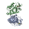
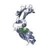
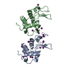
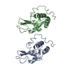
- PDB-6ups: Crystal structure of the deubiquitylase domain from the Orientia ... -
+ Open data

- Basic information
| Entry | Database: PDB / ID: 6ups | ||||||||||||
|---|---|---|---|---|---|---|---|---|---|---|---|---|---|
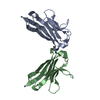
| Title | Crystal structure of the deubiquitylase domain from the Orientia tsutsugamushi protein OTT_1962 (OtDUB) | ||||||||||||
 Components Components | ULP_PROTEASE domain-containing protein | ||||||||||||
 Keywords Keywords |  HYDROLASE / deubiquitylase / Orientia / CE clan HYDROLASE / deubiquitylase / Orientia / CE clan | ||||||||||||
| Function / homology | Ubiquitin-like protease family profile. / Ulp1 protease family, C-terminal catalytic domain / Ulp1 protease family, C-terminal catalytic domain / cysteine-type peptidase activity / Papain-like cysteine peptidase superfamily / proteolysis / Ubiquitin-like protease family profile domain-containing protein Function and homology information Function and homology information | ||||||||||||
| Biological species |  Orientia tsutsugamushi (bacteria) Orientia tsutsugamushi (bacteria) | ||||||||||||
| Method | X-RAY DIFFRACTION / SYNCHROTRON / SAD / Resolution: 2 Å | ||||||||||||
 Authors Authors | Ronau, J.A. / Lim, C.S. / Xiong, Y. | ||||||||||||
| Funding support |  United States, 3items United States, 3items
| ||||||||||||
 Citation Citation | Journal: Nat Commun / Year: 2020 Title: A deubiquitylase with an unusually high-affinity ubiquitin-binding domain from the scrub typhus pathogen Orientia tsutsugamushi. Authors: Berk, J.M. / Lim, C. / Ronau, J.A. / Chaudhuri, A. / Chen, H. / Beckmann, J.F. / Loria, J.P. / Xiong, Y. / Hochstrasser, M. | ||||||||||||
| History |
|
- Structure visualization
| Structure viewer | Molecule: MolmilJmol/JSmol |
|---|
- Downloads & links
-Download
| PDBx/mmCIF format | 6ups.cif.gz | 107.5 KB | Display | PDBx/mmCIF format |
|---|---|---|---|---|
| PDB format | pdb6ups.ent.gz | 86.5 KB | Display | PDB format |
| PDBx/mmJSON format | 6ups.json.gz | Tree view | PDBx/mmJSON format | |
| Others |  Other downloads Other downloads |
-Validation report
| Arichive directory | https://data.pdbj.org/pub/pdb/validation_reports/up/6upsftp://data.pdbj.org/pub/pdb/validation_reports/up/6ups | HTTPS FTP |
|---|
-Related structure data


-Links
 PDBj
PDBj- Assembly
| Deposited unit | 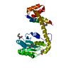
| ||||||||
|---|---|---|---|---|---|---|---|---|---|
| 1 |
| ||||||||
| Unit cell |
|
-Components
| #1: Protein | Mass: 29597.367 Da / Num. of mol.: 1 / Fragment: deubiquitylase domain Source method: isolated from a genetically manipulated source Source: (gene. exp.) Orientia tsutsugamushi (strain Ikeda) (bacteria)Strain: Ikeda / Gene: OTT_1962 / Production host: Escherichia coli (E. coli)References: UniProt: B3CVM3, Hydrolases; Acting on peptide bonds (peptidases); Cysteine endopeptidases |
|---|---|
| #2: Water | ChemComp-HOH / Water Mass: 18.015 Da / Num. of mol.: 80 / Source method: isolated from a natural source / Formula: H2O Mass: 18.015 Da / Num. of mol.: 80 / Source method: isolated from a natural source / Formula: H2O |
| Has ligand of interest | N |
-Experimental details
-Experiment
| Experiment | Method: X-RAY DIFFRACTION / Number of used crystals: 1 |
|---|
- Sample preparation
| Crystal | Density Matthews: 2.5 Å3/Da / Density % sol: 50.76 % |
|---|---|
| Crystal grow | Temperature: 298 K / Method: vapor diffusion, sitting drop / pH: 7.5 / Details: 0.2 M ammonium iodide, 20% (w/v) PEG 3350 |
-Data collection
| Diffraction | Mean temperature: 100 K / Serial crystal experiment: N |
|---|---|
| Diffraction source | Source: SYNCHROTRON / Site: APS / Beamline: 21-ID-E / Wavelength: 0.98 Å |
| Detector | Type: ADSC QUANTUM 315 / Detector: CCD / Date: Jun 10, 2017 |
| Radiation | Monochromator: SI(111) / Protocol: SINGLE WAVELENGTH / Monochromatic (M) / Laue (L): M / Scattering type: x-ray |
| Radiation wavelength | Wavelength: 0.98 Å / Relative weight: 1 |
| Reflection | Resolution: 2→50 Å / Num. obs: 18794 / % possible obs: 91 % / Redundancy: 4.3 % / Rmerge(I) obs: 0.144 / Net I/σ(I): 10.6 |
| Reflection shell | Resolution: 2→2.07 Å / Redundancy: 4 % / Rmerge(I) obs: 0.825 / Mean I/σ(I) obs: 1.6 / Num. unique obs: 1895 / % possible all: 93.7 |
- Processing
| Software |
| ||||||||||||||||||||||||||||||||||||||||||||||||||||||||||||
|---|---|---|---|---|---|---|---|---|---|---|---|---|---|---|---|---|---|---|---|---|---|---|---|---|---|---|---|---|---|---|---|---|---|---|---|---|---|---|---|---|---|---|---|---|---|---|---|---|---|---|---|---|---|---|---|---|---|---|---|---|---|
| Refinement | Method to determine structure: SAD / Resolution: 2→34.67 Å / Cor.coef. Fo:Fc: 0.95 / Cor.coef. Fo:Fc free: 0.935 / SU B: 11.782 / SU ML: 0.145 / Cross valid method: THROUGHOUT / σ(F): 0 / ESU R: 0.195 / ESU R Free: 0.165 Details: U VALUES : WITH TLS ADDED HYDROGENS HAVE BEEN ADDED IN THE RIDING POSITIONS
| ||||||||||||||||||||||||||||||||||||||||||||||||||||||||||||
| Solvent computation | Ion probe radii: 0.8 Å / Shrinkage radii: 0.8 Å / VDW probe radii: 1.2 Å | ||||||||||||||||||||||||||||||||||||||||||||||||||||||||||||
| Displacement parameters | Biso max: 105.93 Å2 / Biso mean: 38.339 Å2 / Biso min: 17.35 Å2
| ||||||||||||||||||||||||||||||||||||||||||||||||||||||||||||
| Refinement step | Cycle: final / Resolution: 2→34.67 Å
| ||||||||||||||||||||||||||||||||||||||||||||||||||||||||||||
| Refine LS restraints |
| ||||||||||||||||||||||||||||||||||||||||||||||||||||||||||||
| LS refinement shell | Resolution: 2→2.052 Å / Rfactor Rfree error: 0 / Total num. of bins used: 20
| ||||||||||||||||||||||||||||||||||||||||||||||||||||||||||||
| Refinement TLS params. | Method: refined / Origin x: 39.76 Å / Origin y: 2.523 Å / Origin z: 28.79 Å
|
