 Movie
Movie Controller

[English] 日本語
 Yorodumi
- PDB-6ume: Crystal structure of human GAC in complex with inhibitor UPGL00012 -
+ Open data

- Basic information
| Entry | Database: PDB / ID: 6ume | ||||||||||||
|---|---|---|---|---|---|---|---|---|---|---|---|---|---|
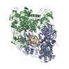
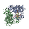
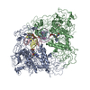
| Title | Crystal structure of human GAC in complex with inhibitor UPGL00012 | ||||||||||||
 Components Components | Glutaminase kidney isoform, mitochondrial | ||||||||||||
 Keywords Keywords | HYDROLASE/HYDROLASE INHIBITOR /  inhibitor / complex / HYDROLASE / HYDROLASE-HYDROLASE INHIBITOR complex inhibitor / complex / HYDROLASE / HYDROLASE-HYDROLASE INHIBITOR complex | ||||||||||||
| Function / homology |  Function and homology information Function and homology informationglutamine catabolic process / regulation of respiratory gaseous exchange by nervous system process / glutamate biosynthetic process / Glutamate and glutamine metabolism / intracellular glutamate homeostasis / Glutamate Neurotransmitter Release Cycle / glutaminase / glutaminase activity / suckling behavior / TP53 Regulates Metabolic Genes ...glutamine catabolic process / regulation of respiratory gaseous exchange by nervous system process / glutamate biosynthetic process / Glutamate and glutamine metabolism / intracellular glutamate homeostasis / Glutamate Neurotransmitter Release Cycle / glutaminase / glutaminase activity / suckling behavior / TP53 Regulates Metabolic Genes / chemical synaptic transmission / protein homotetramerization / mitochondrial matrix / synapse / mitochondrion / cytosolSimilarity search - Function | ||||||||||||
| Biological species |  Homo sapiens (human) Homo sapiens (human) | ||||||||||||
| Method | X-RAY DIFFRACTION / SYNCHROTRON / MOLECULAR REPLACEMENT / Resolution: 2.9 Å | ||||||||||||
 Authors Authors | Huang, Q. / Cerione, R.A. | ||||||||||||
| Funding support |  United States, 3items United States, 3items
| ||||||||||||
 Citation Citation | Journal: To Be Published Title: Crystal structure of human GAC in complex with inhibitor UPGL00012 Authors: Huang, Q. / Cerione, R.A. #1: Journal: J. Biol. Chem. / Year: 2018Title: Characterization of the interactions of potent allosteric inhibitors with glutaminase C, a key enzyme in cancer cell glutamine metabolism. Authors: Huang, Q. / Stalnecker, C.A. / Zhang, C. / McDermott, L.A. / Iyer, P. / O'Neill, J. / Reimer, S. / Cerione, R.A. / Katt, W.P. #2: Journal: Bioorg. Med. Chem. / Year: 2016Title: Design and evaluation of novel glutaminase inhibitors. Authors: McDermott, L.A. / Iyer, P. / Vernetti, L. / Reimer, S. / Sun, J. / Boby, M. / Yang, T. / Fioravanti, M. / O'Neill, J. / Wang, L. / Drakes, D. / Katt, W.P. / Huang, Q. / Cerione, R.A. #3: Journal: J. Biol. Chem. / Year: 2016Title: Mechanistic Basis of Glutaminase Activation: A KEY ENZYME THAT PROMOTES GLUTAMINE METABOLISM IN CANCER CELLS. Authors: Li, Y. / Erickson, J.W. / Stalnecker, C.A. / Katt, W.P. / Huang, Q. / Cerione, R.A. / Ramachandran, S. | ||||||||||||
| History |
|
- Structure visualization
| Structure viewer | Molecule: MolmilJmol/JSmol |
|---|
- Downloads & links
-Download
| PDBx/mmCIF format | 6ume.cif.gz | 771.8 KB | Display | PDBx/mmCIF format |
|---|---|---|---|---|
| PDB format | pdb6ume.ent.gz | 540.1 KB | Display | PDB format |
| PDBx/mmJSON format | 6ume.json.gz | Tree view | PDBx/mmJSON format | |
| Others |  Other downloads Other downloads |
-Validation report
| Arichive directory | https://data.pdbj.org/pub/pdb/validation_reports/um/6umeftp://data.pdbj.org/pub/pdb/validation_reports/um/6ume | HTTPS FTP |
|---|
-Related structure data
| Related structure data |  6ujgC  6ul9C  6umcC  6umdC  6umfC  5d3oS S: Starting model for refinement C: citing same article ( |
|---|---|
| Similar structure data |


-Links
 PDBj
PDBj
- Assembly
| Deposited unit | 
| |||||||||||||||||||||||||||||||||||
|---|---|---|---|---|---|---|---|---|---|---|---|---|---|---|---|---|---|---|---|---|---|---|---|---|---|---|---|---|---|---|---|---|---|---|---|---|
| 1 |
| |||||||||||||||||||||||||||||||||||
| Unit cell |
| |||||||||||||||||||||||||||||||||||
| Noncrystallographic symmetry (NCS) | NCS domain:
NCS domain segments: Ens-ID: 1 / Beg auth comp-ID: PRO / Beg label comp-ID: PRO / End auth comp-ID: ARG / End label comp-ID: ARG / Auth seq-ID: 137 - 544 / Label seq-ID: 66 - 473
|
-Components
| #1: Protein | Mass: 58023.996 Da / Num. of mol.: 4 Source method: isolated from a genetically manipulated source Source: (gene. exp.) Homo sapiens (human) / Gene: GLS, GLS1, KIAA0838 / Production host:  Escherichia coli (E. coli) / References: UniProt: O94925, glutaminase Escherichia coli (E. coli) / References: UniProt: O94925, glutaminase#2: Chemical |   Mass: 536.632 Da / Num. of mol.: 2 / Source method: obtained synthetically / Formula: C23H24N10O2S2 / Feature type: SUBJECT OF INVESTIGATION Mass: 536.632 Da / Num. of mol.: 2 / Source method: obtained synthetically / Formula: C23H24N10O2S2 / Feature type: SUBJECT OF INVESTIGATIONHas ligand of interest | Y | |
|---|
-Experimental details
-Experiment
| Experiment | Method: X-RAY DIFFRACTION / Number of used crystals: 1 |
|---|
- Sample preparation
| Crystal | Density Matthews: 2.69 Å3/Da / Density % sol: 54.34 % |
|---|---|
| Crystal grow | Temperature: 293 K / Method: vapor diffusion, hanging drop / pH: 8 / Details: 12% PEG6000, 1.0 M LiCl pH8.0 / PH range: 8-8.5 |
-Data collection
| Diffraction | Mean temperature: 100 K / Serial crystal experiment: N |
|---|---|
| Diffraction source | Source: SYNCHROTRON / Site: CHESS / Beamline: A1 / Wavelength: 0.98 Å |
| Detector | Type: ADSC QUANTUM 210 / Detector: CCD / Date: May 29, 2015 |
| Radiation | Protocol: SINGLE WAVELENGTH / Monochromatic (M) / Laue (L): M / Scattering type: x-ray |
| Radiation wavelength | Wavelength: 0.98 Å / Relative weight: 1 |
| Reflection | Resolution: 2.8→50 Å / Num. obs: 121885 / % possible obs: 99.2 % / Redundancy: 3.7 % / Biso Wilson estimate: 57.88 Å2 / Rrim(I) all: 0.179 / Rsym value: 0.167 / Net I/σ(I): 8.89 |
| Reflection shell | Resolution: 2.8→2.85 Å / Redundancy: 3.1 % / Mean I/σ(I) obs: 1.05 / Num. unique obs: 5554 / CC1/2: 0.811 / Rsym value: 0.47 / % possible all: 91.2 |
- Processing
| Software |
| ||||||||||||||||||||||||||||||||||||||||||||||||||||||||||||||||||||||||||||||||||||||||||||||||||
|---|---|---|---|---|---|---|---|---|---|---|---|---|---|---|---|---|---|---|---|---|---|---|---|---|---|---|---|---|---|---|---|---|---|---|---|---|---|---|---|---|---|---|---|---|---|---|---|---|---|---|---|---|---|---|---|---|---|---|---|---|---|---|---|---|---|---|---|---|---|---|---|---|---|---|---|---|---|---|---|---|---|---|---|---|---|---|---|---|---|---|---|---|---|---|---|---|---|---|---|
| Refinement | Method to determine structure: MOLECULAR REPLACEMENT Starting model: 5d3o Resolution: 2.9→24.46 Å / SU ML: 0.3579 / Cross valid method: FREE R-VALUE / σ(F): 1.34 / Phase error: 39.7949
| ||||||||||||||||||||||||||||||||||||||||||||||||||||||||||||||||||||||||||||||||||||||||||||||||||
| Solvent computation | Shrinkage radii: 0.9 Å / VDW probe radii: 1.11 Å | ||||||||||||||||||||||||||||||||||||||||||||||||||||||||||||||||||||||||||||||||||||||||||||||||||
| Displacement parameters | Biso mean: 71.57 Å2 | ||||||||||||||||||||||||||||||||||||||||||||||||||||||||||||||||||||||||||||||||||||||||||||||||||
| Refinement step | Cycle: LAST / Resolution: 2.9→24.46 Å
| ||||||||||||||||||||||||||||||||||||||||||||||||||||||||||||||||||||||||||||||||||||||||||||||||||
| Refine LS restraints |
| ||||||||||||||||||||||||||||||||||||||||||||||||||||||||||||||||||||||||||||||||||||||||||||||||||
| LS refinement shell |
| ||||||||||||||||||||||||||||||||||||||||||||||||||||||||||||||||||||||||||||||||||||||||||||||||||
| Refinement TLS params. | Method: refined / Origin x: -54.4876097038 Å / Origin y: 37.277010657 Å / Origin z: -31.2962188203 Å
| ||||||||||||||||||||||||||||||||||||||||||||||||||||||||||||||||||||||||||||||||||||||||||||||||||
| Refinement TLS group | Selection details: all |
