 Movie
Movie Controller

[English] 日本語
 Yorodumi
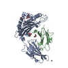
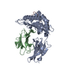
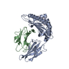
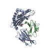
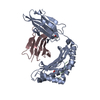
- PDB-6ulk: Molecular basis for tumor infiltrating TCR recognition of hotspot... -
+ Open data

- Basic information
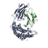
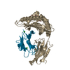
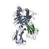
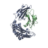
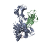
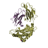
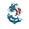
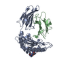
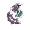
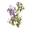
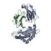
| Entry | Database: PDB / ID: 6ulk | ||||||
|---|---|---|---|---|---|---|---|
| Title | Molecular basis for tumor infiltrating TCR recognition of hotspot KRAS-G12D mutation | ||||||
 Components Components |
| ||||||
 Keywords Keywords |  IMMUNE SYSTEM / HLA-C Neoantigen KRAS IMMUNE SYSTEM / HLA-C Neoantigen KRAS | ||||||
| Function / homology |  Function and homology information Function and homology informationmyoblast differentiation / Signaling by RAS GAP mutants / Signaling by RAS GTPase mutants / Activation of RAS in B cells / tertiary granule membrane / RAS signaling downstream of NF1 loss-of-function variants / SOS-mediated signalling / antigen processing and presentation / Activated NTRK3 signals through RAS / Activated NTRK2 signals through RAS ...myoblast differentiation / Signaling by RAS GAP mutants / Signaling by RAS GTPase mutants / Activation of RAS in B cells / tertiary granule membrane / RAS signaling downstream of NF1 loss-of-function variants / SOS-mediated signalling / antigen processing and presentation / Activated NTRK3 signals through RAS / Activated NTRK2 signals through RAS / SHC1 events in ERBB4 signaling / Signalling to RAS / SHC-related events triggered by IGF1R / Activated NTRK2 signals through FRS2 and FRS3 / Estrogen-stimulated signaling through PRKCZ / SHC-mediated cascade:FGFR3 / MET activates RAS signaling / PTK6 Regulates RHO GTPases, RAS GTPase and MAP kinases / Signaling by PDGFRA transmembrane, juxtamembrane and kinase domain mutants / Signaling by PDGFRA extracellular domain mutants / SHC-mediated cascade:FGFR2 / SHC-mediated cascade:FGFR4 / Signaling by FGFR4 in disease / SHC-mediated cascade:FGFR1 / Erythropoietin activates RAS / FRS-mediated FGFR3 signaling / Signaling by FLT3 ITD and TKD mutants / FRS-mediated FGFR2 signaling / FRS-mediated FGFR4 signaling / Signaling by FGFR3 in disease / FRS-mediated FGFR1 signaling / p38MAPK events / Tie2 Signaling / positive regulation of endothelial cell proliferation / Signaling by FGFR2 in disease / GRB2 events in EGFR signaling / SHC1 events in EGFR signaling / EGFR Transactivation by Gastrin / Signaling by FLT3 fusion proteins / FLT3 Signaling / Signaling by FGFR1 in disease / Ras activation upon Ca2+ influx through NMDA receptor / GRB2 events in ERBB2 signaling / NCAM signaling for neurite out-growth / CD209 (DC-SIGN) signaling / SHC1 events in ERBB2 signaling / Downstream signal transduction / Constitutive Signaling by Overexpressed ERBB2 / Insulin receptor signalling cascade / small monomeric GTPase / G protein activity / Signaling by phosphorylated juxtamembrane, extracellular and kinase domain KIT mutants / positive regulation of ferrous iron binding / positive regulation of transferrin receptor binding / VEGFR2 mediated cell proliferation / positive regulation of receptor binding / early endosome lumen / Nef mediated downregulation of MHC class I complex cell surface expression / DAP12 interactions / negative regulation of receptor binding / lumenal side of endoplasmic reticulum membrane / Endosomal/Vacuolar pathway / FCERI mediated MAPK activation / Antigen Presentation: Folding, assembly and peptide loading of class I MHC / Signaling by ERBB2 TMD/JMD mutants / RAF activation / Signaling by high-kinase activity BRAF mutants / cellular response to iron(III) ion / antigen processing and presentation of exogenous protein antigen via MHC class Ib, TAP-dependent / Constitutive Signaling by EGFRvIII / negative regulation of forebrain neuron differentiation / MAP2K and MAPK activation / Signaling by ERBB2 ECD mutants / ER to Golgi transport vesicle membrane / regulation of erythrocyte differentiation / peptide antigen assembly with MHC class I protein complex / response to molecule of bacterial origin / regulation of iron ion transport / MHC class I peptide loading complex / Signaling by ERBB2 KD Mutants / Signaling by SCF-KIT / HFE-transferrin receptor complex / T cell mediated cytotoxicity / cellular response to iron ion / antigen processing and presentation of endogenous peptide antigen via MHC class I / positive regulation of T cell cytokine production / MHC class I protein complex / multicellular organismal-level iron ion homeostasis / negative regulation of neurogenesis / peptide antigen assembly with MHC class II protein complex / positive regulation of receptor-mediated endocytosis / positive regulation of T cell mediated cytotoxicity / MHC class II protein complex / cellular response to nicotine / Regulation of RAS by GAPs / Negative regulation of MAPK pathway / recycling endosome membrane / RAS processing / specific granule lumen / phagocytic vesicle membraneSimilarity search - Function | ||||||
| Biological species |  Homo sapiens (human) Homo sapiens (human) | ||||||
| Method | X-RAY DIFFRACTION / SYNCHROTRON / MOLECULAR REPLACEMENT / Resolution: 1.9 Å | ||||||
 Authors Authors | Sun, P.D. / Sim, M.J.W. | ||||||
 Citation Citation | Journal: Proc.Natl.Acad.Sci.USA / Year: 2020 Title: High-affinity oligoclonal TCRs define effective adoptive T cell therapy targeting mutant KRAS-G12D. Authors: Sim, M.J.W. / Lu, J. / Spencer, M. / Hopkins, F. / Tran, E. / Rosenberg, S.A. / Long, E.O. / Sun, P.D. | ||||||
| History |
|
- Structure visualization
| Structure viewer | Molecule: MolmilJmol/JSmol |
|---|
- Downloads & links
-Download
| PDBx/mmCIF format | 6ulk.cif.gz | 208.5 KB | Display | PDBx/mmCIF format |
|---|---|---|---|---|
| PDB format | pdb6ulk.ent.gz | 138.5 KB | Display | PDB format |
| PDBx/mmJSON format | 6ulk.json.gz | Tree view | PDBx/mmJSON format | |
| Others |  Other downloads Other downloads |
-Validation report
| Arichive directory | https://data.pdbj.org/pub/pdb/validation_reports/ul/6ulkftp://data.pdbj.org/pub/pdb/validation_reports/ul/6ulk | HTTPS FTP |
|---|
-Related structure data
| Related structure data |  6uliC  6ulnC  6ulrC  6uonC 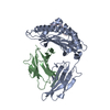 4nt6S S: Starting model for refinement C: citing same article ( |
|---|---|
| Similar structure data |


-Links
 PDBj
PDBj


- Assembly
| Deposited unit | 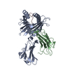
| ||||||||||||
|---|---|---|---|---|---|---|---|---|---|---|---|---|---|
| 1 |
| ||||||||||||
| Unit cell |
| ||||||||||||
| Components on special symmetry positions |
|
-Components
| #1: Protein | Mass: 31837.107 Da / Num. of mol.: 1 Source method: isolated from a genetically manipulated source Source: (gene. exp.) Homo sapiens (human) / Gene: HLA-Cw, HLA-C / Production host:  Escherichia coli (E. coli) / References: UniProt: C1K0Y1 Escherichia coli (E. coli) / References: UniProt: C1K0Y1 |
|---|---|
| #2: Protein | Beta-2 microglobulin Mass: 11879.356 Da / Num. of mol.: 1 Source method: isolated from a genetically manipulated source Source: (gene. exp.) Homo sapiens (human) / Gene: B2M, CDABP0092, HDCMA22P / Production host: Escherichia coli (E. coli) / References: UniProt: P61769 |
| #3: Protein/peptide | Mass: 874.960 Da / Num. of mol.: 1 / Mutation: G12D / Source method: obtained synthetically / Source: (synth.) Homo sapiens (human) / References: UniProt: P01111 |
| #4: Water | ChemComp-HOH / Water Mass: 18.015 Da / Num. of mol.: 148 / Source method: isolated from a natural source / Formula: H2O Mass: 18.015 Da / Num. of mol.: 148 / Source method: isolated from a natural source / Formula: H2O |
-Experimental details
-Experiment
| Experiment | Method: X-RAY DIFFRACTION / Number of used crystals: 1 |
|---|
- Sample preparation
| Crystal | Density Matthews: 2.22 Å3/Da / Density % sol: 44.49 % |
|---|---|
| Crystal grow | Temperature: 293 K / Method: vapor diffusion, hanging drop Details: 0.1 M Bis-Tris pH 6.5, 0.05M CaCl2 dihydrate, 30% PEG MME 500 |
-Data collection
| Diffraction | Mean temperature: 100 K / Serial crystal experiment: N |
|---|---|
| Diffraction source | Source: SYNCHROTRON / Site: APS  / Beamline: 22-ID / Wavelength: 1 Å / Beamline: 22-ID / Wavelength: 1 Å |
| Detector | Type: DECTRIS EIGER X 16M / Detector: PIXEL / Date: Mar 3, 2017 |
| Radiation | Protocol: SINGLE WAVELENGTH / Monochromatic (M) / Laue (L): M / Scattering type: x-ray |
| Radiation wavelength | Wavelength: 1 Å / Relative weight: 1 |
| Reflection | Resolution: 1.88→33.44 Å / Num. obs: 30790 / % possible obs: 99.5 % / Redundancy: 5.3 % / Biso Wilson estimate: 21.93 Å2 / Rmerge(I) obs: 0.112 / Net I/σ(I): 21.8 |
| Reflection shell | Resolution: 1.88→1.9 Å / Rmerge(I) obs: 0.395 / Num. unique obs: 1574 |
- Processing
| Software |
| ||||||||||||||||||||||||||||||||||||||||||||||||||||||||||||||||||||||||||||||||||||
|---|---|---|---|---|---|---|---|---|---|---|---|---|---|---|---|---|---|---|---|---|---|---|---|---|---|---|---|---|---|---|---|---|---|---|---|---|---|---|---|---|---|---|---|---|---|---|---|---|---|---|---|---|---|---|---|---|---|---|---|---|---|---|---|---|---|---|---|---|---|---|---|---|---|---|---|---|---|---|---|---|---|---|---|---|---|
| Refinement | Method to determine structure: MOLECULAR REPLACEMENT Starting model: 4NT6 Resolution: 1.9→33.44 Å / SU ML: 0.2297 / Cross valid method: FREE R-VALUE / σ(F): 1.38 / Phase error: 29.3532 Stereochemistry target values: GeoStd + Monomer Library + CDL v1.2
| ||||||||||||||||||||||||||||||||||||||||||||||||||||||||||||||||||||||||||||||||||||
| Solvent computation | Shrinkage radii: 0.9 Å / VDW probe radii: 1.11 Å / Solvent model: FLAT BULK SOLVENT MODEL | ||||||||||||||||||||||||||||||||||||||||||||||||||||||||||||||||||||||||||||||||||||
| Displacement parameters | Biso mean: 31.56 Å2 | ||||||||||||||||||||||||||||||||||||||||||||||||||||||||||||||||||||||||||||||||||||
| Refinement step | Cycle: LAST / Resolution: 1.9→33.44 Å
| ||||||||||||||||||||||||||||||||||||||||||||||||||||||||||||||||||||||||||||||||||||
| Refine LS restraints |
| ||||||||||||||||||||||||||||||||||||||||||||||||||||||||||||||||||||||||||||||||||||
| LS refinement shell |
| ||||||||||||||||||||||||||||||||||||||||||||||||||||||||||||||||||||||||||||||||||||
| Refinement TLS params. | Method: refined / Origin x: 18.2435334974 Å / Origin y: 1.68102160926 Å / Origin z: 23.087645579 Å
| ||||||||||||||||||||||||||||||||||||||||||||||||||||||||||||||||||||||||||||||||||||
| Refinement TLS group | Selection details: all |
