 Movie
Movie Controller


+ Open data

- Basic information
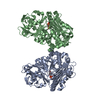
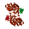
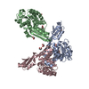
| Entry | Database: PDB / ID: 6ui5 | |||||||||
|---|---|---|---|---|---|---|---|---|---|---|
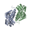
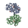
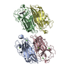
| Title | Tmn9 in complex with cofactor FAD | |||||||||
 Components Components | 2-polyprenyl-6-methoxyphenol hydroxylase-like FAD-dependent oxidoreductase | |||||||||
 Keywords Keywords |  OXIDOREDUCTASE / Complex / cyclase / UNKNOWN FUNCTION OXIDOREDUCTASE / Complex / cyclase / UNKNOWN FUNCTION | |||||||||
| Function / homology | FAD-binding domain / FAD binding domain / FAD binding / monooxygenase activity / FAD/NAD(P)-binding domain superfamily / FLAVIN-ADENINE DINUCLEOTIDE / 2-polyprenyl-6-methoxyphenol hydroxylase-like FAD-dependent oxidoreductase / Putative FAD-dependent monooxygenase Function and homology information Function and homology information | |||||||||
| Biological species |  Streptomyces sp. NRRL 11266 (bacteria) Streptomyces sp. NRRL 11266 (bacteria) | |||||||||
| Method | X-RAY DIFFRACTION / SYNCHROTRON / MOLECULAR REPLACEMENT / Resolution: 2.4 Å | |||||||||
 Authors Authors | Paiva, F.C.R. / Little, R. / Leadlay, P. / Dias, M.V.B. | |||||||||
| Funding support |  Brazil, 2items Brazil, 2items
| |||||||||
 Citation Citation | Journal: To Be Published Title: Tmn9 in complex with cofactor FAD Authors: Paiva, F.C.R. / Little, R. / Leadlay, P. / Dias, M.V.B. | |||||||||
| History |
|
- Structure visualization
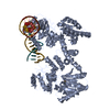
| Structure viewer | Molecule: MolmilJmol/JSmol |
|---|
- Downloads & links
-Download
| PDBx/mmCIF format | 6ui5.cif.gz | 402.4 KB | Display | PDBx/mmCIF format |
|---|---|---|---|---|
| PDB format | pdb6ui5.ent.gz | 330.5 KB | Display | PDB format |
| PDBx/mmJSON format | 6ui5.json.gz | Tree view | PDBx/mmJSON format | |
| Others |  Other downloads Other downloads |
-Validation report
| Arichive directory | https://data.pdbj.org/pub/pdb/validation_reports/ui/6ui5ftp://data.pdbj.org/pub/pdb/validation_reports/ui/6ui5 | HTTPS FTP |
|---|
-Related structure data
| Related structure data | 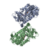 5xgvS S: Starting model for refinement |
|---|---|
| Similar structure data |

-Links
 PDBj
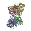
PDBj- Assembly
| Deposited unit | 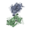
| ||||||||
|---|---|---|---|---|---|---|---|---|---|
| 1 |
| ||||||||
| Unit cell |
|
-Components
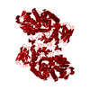
| #1: Protein | Mass: 54334.340 Da / Num. of mol.: 2 Source method: isolated from a genetically manipulated source Source: (gene. exp.) Streptomyces sp. NRRL 11266 (bacteria) / Production host: Escherichia coli BL21(DE3) (bacteria) / References: UniProt: A0A2M8ZYZ6, UniProt: Q1MX79*PLUS#2: Chemical | Flavin adenine dinucleotide  Mass: 785.550 Da / Num. of mol.: 2 / Source method: obtained synthetically / Formula: C27H33N9O15P2 / Comment: FAD*YM Mass: 785.550 Da / Num. of mol.: 2 / Source method: obtained synthetically / Formula: C27H33N9O15P2 / Comment: FAD*YM#3: Water | ChemComp-HOH / | Water Mass: 18.015 Da / Num. of mol.: 38 / Source method: isolated from a natural source / Formula: H2O Mass: 18.015 Da / Num. of mol.: 38 / Source method: isolated from a natural source / Formula: H2OHas ligand of interest | N | |
|---|
-Experimental details
-Experiment
| Experiment | Method: X-RAY DIFFRACTION / Number of used crystals: 1 |
|---|
- Sample preparation
| Crystal | Density Matthews: 2.5 Å3/Da / Density % sol: 50.87 % |
|---|---|
| Crystal grow | Temperature: 291.15 K / Method: vapor diffusion, hanging drop Details: 30% w/v PEG 8000, 100 mM sodium cacodylate pH 6,5, 200 mM sodium acetate |
-Data collection
| Diffraction | Mean temperature: 100 K / Serial crystal experiment: N | ||||||||||||||||||||||||||||||
|---|---|---|---|---|---|---|---|---|---|---|---|---|---|---|---|---|---|---|---|---|---|---|---|---|---|---|---|---|---|---|---|
| Diffraction source | Source: SYNCHROTRON / Site: PETRA III, EMBL c/o DESY  / Beamline: P13 (MX1) / Wavelength: 0.976255 Å / Beamline: P13 (MX1) / Wavelength: 0.976255 Å | ||||||||||||||||||||||||||||||
| Detector | Type: DECTRIS PILATUS 6M-F / Detector: PIXEL / Date: Jun 26, 2018 | ||||||||||||||||||||||||||||||
| Radiation | Protocol: SINGLE WAVELENGTH / Monochromatic (M) / Laue (L): M / Scattering type: x-ray | ||||||||||||||||||||||||||||||
| Radiation wavelength | Wavelength: 0.976255 Å / Relative weight: 1 | ||||||||||||||||||||||||||||||
| Reflection | Resolution: 2.26→49.79 Å / Num. obs: 52087 / % possible obs: 99.6 % / Redundancy: 6.3 % / Biso Wilson estimate: 57.47 Å2 / CC1/2: 0.996 / Rmerge(I) obs: 0.078 / Rpim(I) all: 0.034 / Rrim(I) all: 0.085 / Net I/σ(I): 12 / Num. measured all: 328949 / Scaling rejects: 22 | ||||||||||||||||||||||||||||||
| Reflection shell | Diffraction-ID: 1
|
- Processing
| Software |
| ||||||||||||||||||||||||||||||||||||||||||||||||||||||||||||||||||||||||||||||||||||||||||||||||
|---|---|---|---|---|---|---|---|---|---|---|---|---|---|---|---|---|---|---|---|---|---|---|---|---|---|---|---|---|---|---|---|---|---|---|---|---|---|---|---|---|---|---|---|---|---|---|---|---|---|---|---|---|---|---|---|---|---|---|---|---|---|---|---|---|---|---|---|---|---|---|---|---|---|---|---|---|---|---|---|---|---|---|---|---|---|---|---|---|---|---|---|---|---|---|---|---|---|
| Refinement | Method to determine structure: MOLECULAR REPLACEMENT Starting model: 5XGV Resolution: 2.4→49.788 Å / SU ML: 0.4 / Cross valid method: THROUGHOUT / σ(F): 1.34 / Phase error: 30.75
| ||||||||||||||||||||||||||||||||||||||||||||||||||||||||||||||||||||||||||||||||||||||||||||||||
| Solvent computation | Shrinkage radii: 0.9 Å / VDW probe radii: 1.11 Å | ||||||||||||||||||||||||||||||||||||||||||||||||||||||||||||||||||||||||||||||||||||||||||||||||
| Displacement parameters | Biso max: 222.85 Å2 / Biso mean: 83.9249 Å2 / Biso min: 32.1 Å2 | ||||||||||||||||||||||||||||||||||||||||||||||||||||||||||||||||||||||||||||||||||||||||||||||||
| Refinement step | Cycle: final / Resolution: 2.4→49.788 Å
| ||||||||||||||||||||||||||||||||||||||||||||||||||||||||||||||||||||||||||||||||||||||||||||||||
| Refine LS restraints |
| ||||||||||||||||||||||||||||||||||||||||||||||||||||||||||||||||||||||||||||||||||||||||||||||||
| LS refinement shell | Refine-ID: X-RAY DIFFRACTION / Rfactor Rfree error: 0
| ||||||||||||||||||||||||||||||||||||||||||||||||||||||||||||||||||||||||||||||||||||||||||||||||
| Refinement TLS params. | Method: refined / Origin x: 35.2767 Å / Origin y: -11.2119 Å / Origin z: 233.1232 Å
| ||||||||||||||||||||||||||||||||||||||||||||||||||||||||||||||||||||||||||||||||||||||||||||||||
| Refinement TLS group |
|
