 Movie
Movie Controller


+ Open data

- Basic information
| Entry | Database: PDB / ID: 6u8z | |||||||||
|---|---|---|---|---|---|---|---|---|---|---|
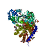
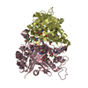
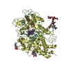
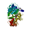
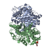
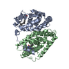
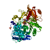
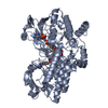
| Title | Crystal Structure of Catalytic Domain of Human Phospholipase D1 | |||||||||
 Components Components | Phospholipase D1,Phospholipase D1 | |||||||||
 Keywords Keywords |  HYDROLASE / Phospholipase D1 / PLD / phosphohistidine / PIP2 binding HYDROLASE / Phospholipase D1 / PLD / phosphohistidine / PIP2 binding | |||||||||
| Function / homology |  Function and homology information: / Synthesis of PG / N-acylphosphatidylethanolamine-specific phospholipase D activity / phospholipase D / phosphatidic acid biosynthetic process / regulation of microvillus assembly / Synthesis of PA / phospholipase D activity / phospholipid catabolic process / cholinergic synapse ...: / Synthesis of PG / N-acylphosphatidylethanolamine-specific phospholipase D activity / phospholipase D / phosphatidic acid biosynthetic process / regulation of microvillus assembly / Synthesis of PA / phospholipase D activity / phospholipid catabolic process / cholinergic synapse / regulation of synaptic vesicle cycle / regulation of vesicle-mediated transport / small GTPase-mediated signal transduction / tertiary granule membrane / CDC42 GTPase cycle / endocytic vesicle / RHOG GTPase cycle / RHOA GTPase cycle / Role of phospholipids in phagocytosis / specific granule membrane / RAC1 GTPase cycle / phosphatidylinositol binding / positive regulation of translation / chemotaxis / late endosome membrane / Ras protein signal transduction / endosome / apical plasma membrane / lysosomal membrane / Golgi membrane / intracellular membrane-bounded organelle / Neutrophil degranulation / endoplasmic reticulum membrane / perinuclear region of cytoplasm / Golgi apparatus / membrane / plasma membrane Function and homology information: / Synthesis of PG / N-acylphosphatidylethanolamine-specific phospholipase D activity / phospholipase D / phosphatidic acid biosynthetic process / regulation of microvillus assembly / Synthesis of PA / phospholipase D activity / phospholipid catabolic process / cholinergic synapse ...: / Synthesis of PG / N-acylphosphatidylethanolamine-specific phospholipase D activity / phospholipase D / phosphatidic acid biosynthetic process / regulation of microvillus assembly / Synthesis of PA / phospholipase D activity / phospholipid catabolic process / cholinergic synapse / regulation of synaptic vesicle cycle / regulation of vesicle-mediated transport / small GTPase-mediated signal transduction / tertiary granule membrane / CDC42 GTPase cycle / endocytic vesicle / RHOG GTPase cycle / RHOA GTPase cycle / Role of phospholipids in phagocytosis / specific granule membrane / RAC1 GTPase cycle / phosphatidylinositol binding / positive regulation of translation / chemotaxis / late endosome membrane / Ras protein signal transduction / endosome / apical plasma membrane / lysosomal membrane / Golgi membrane / intracellular membrane-bounded organelle / Neutrophil degranulation / endoplasmic reticulum membrane / perinuclear region of cytoplasm / Golgi apparatus / membrane / plasma membraneSimilarity search - Function | |||||||||
| Biological species |  Homo sapiens (human) Homo sapiens (human) | |||||||||
| Method | X-RAY DIFFRACTION / SYNCHROTRON / SAD / Resolution: 1.799 Å | |||||||||
 Authors Authors | Bowling, F.Z. / Airola, M.V. | |||||||||
| Funding support |  United States, 2items United States, 2items
| |||||||||
 Citation Citation | Journal: Nat.Chem.Biol. / Year: 2020 Title: Crystal structure of human PLD1 provides insight into activation by PI(4,5)P2and RhoA. Authors: Bowling, F.Z. / Salazar, C.M. / Bell, J.A. / Huq, T.S. / Frohman, M.A. / Airola, M.V. | |||||||||
| History |
|
- Structure visualization
| Structure viewer | Molecule: MolmilJmol/JSmol |
|---|
- Downloads & links
-Download
| PDBx/mmCIF format | 6u8z.cif.gz | 372.8 KB | Display | PDBx/mmCIF format |
|---|---|---|---|---|
| PDB format | pdb6u8z.ent.gz | 320.8 KB | Display | PDB format |
| PDBx/mmJSON format | 6u8z.json.gz | Tree view | PDBx/mmJSON format | |
| Others |  Other downloads Other downloads |
-Validation report
| Arichive directory | https://data.pdbj.org/pub/pdb/validation_reports/u8/6u8zftp://data.pdbj.org/pub/pdb/validation_reports/u8/6u8z | HTTPS FTP |
|---|
-Related structure data
| Similar structure data |
|---|


-Links
 PDBj
PDBj


- Assembly
| Deposited unit | 
| ||||||||
|---|---|---|---|---|---|---|---|---|---|
| 1 |
| ||||||||
| Unit cell |
|
-Components
| #1: Protein | Mass: 69671.414 Da / Num. of mol.: 1 / Fragment: UNP residues 330-500,643-1074 Source method: isolated from a genetically manipulated source Source: (gene. exp.) Homo sapiens (human) / Gene: PLD1 / Plasmid: pFastbac Htb / Cell line (production host): Sf9 / Production host:   Spodoptera frugiperda (fall armyworm) / References: UniProt: Q13393, phospholipase D Spodoptera frugiperda (fall armyworm) / References: UniProt: Q13393, phospholipase D |
|---|---|
| #2: Chemical | ChemComp-PO3 / Phosphite ester  Mass: 78.972 Da / Num. of mol.: 1 / Source method: obtained synthetically / Formula: PO3 Mass: 78.972 Da / Num. of mol.: 1 / Source method: obtained synthetically / Formula: PO3 |
| #3: Water | ChemComp-HOH / Water Mass: 18.015 Da / Num. of mol.: 474 / Source method: isolated from a natural source / Formula: H2O Mass: 18.015 Da / Num. of mol.: 474 / Source method: isolated from a natural source / Formula: H2O |
| Has ligand of interest | N |
-Experimental details
-Experiment
| Experiment | Method: X-RAY DIFFRACTION / Number of used crystals: 1 |
|---|
- Sample preparation
| Crystal | Density Matthews: 2.43 Å3/Da / Density % sol: 49.43 % |
|---|---|
| Crystal grow | Temperature: 298 K / Method: vapor diffusion, hanging drop / pH: 5.3 / Details: 0.2 M ammonium citrate, 15% PEG3350, pH 5.3 |
-Data collection
| Diffraction | Mean temperature: 193 K / Serial crystal experiment: N | ||||||||||||||||||||||||
|---|---|---|---|---|---|---|---|---|---|---|---|---|---|---|---|---|---|---|---|---|---|---|---|---|---|
| Diffraction source | Source: SYNCHROTRON / Site: NSLS / Beamline: X17B1 / Wavelength: 0.97933 Å | ||||||||||||||||||||||||
| Detector | Type: DECTRIS EIGER X 16M / Detector: PIXEL / Date: Nov 14, 2017 | ||||||||||||||||||||||||
| Radiation | Protocol: SINGLE WAVELENGTH / Monochromatic (M) / Laue (L): M / Scattering type: x-ray | ||||||||||||||||||||||||
| Radiation wavelength | Wavelength: 0.97933 Å / Relative weight: 1 | ||||||||||||||||||||||||
| Reflection | Resolution: 1.799→79.88 Å / Num. obs: 64302 / % possible obs: 99 % / Redundancy: 8.7 % / Biso Wilson estimate: 30.221 Å2 / Rpim(I) all: 0.053 / Rrim(I) all: 0.158 / Net I/σ(I): 7 | ||||||||||||||||||||||||
| Reflection shell | Diffraction-ID: 1
|
- Processing
| Software |
| ||||||||||||||||||||||||||||||||||||||||||||||||||||||||||||||||||||||||||||||||||||
|---|---|---|---|---|---|---|---|---|---|---|---|---|---|---|---|---|---|---|---|---|---|---|---|---|---|---|---|---|---|---|---|---|---|---|---|---|---|---|---|---|---|---|---|---|---|---|---|---|---|---|---|---|---|---|---|---|---|---|---|---|---|---|---|---|---|---|---|---|---|---|---|---|---|---|---|---|---|---|---|---|---|---|---|---|---|
| Refinement | Method to determine structure: SAD / Resolution: 1.799→54.759 Å / SU ML: 0.19 / Cross valid method: THROUGHOUT / σ(F): 0 / Phase error: 25.27
| ||||||||||||||||||||||||||||||||||||||||||||||||||||||||||||||||||||||||||||||||||||
| Solvent computation | Shrinkage radii: 0.9 Å / VDW probe radii: 1.11 Å | ||||||||||||||||||||||||||||||||||||||||||||||||||||||||||||||||||||||||||||||||||||
| Displacement parameters | Biso max: 140.43 Å2 / Biso mean: 49.9468 Å2 / Biso min: 19.48 Å2 | ||||||||||||||||||||||||||||||||||||||||||||||||||||||||||||||||||||||||||||||||||||
| Refinement step | Cycle: final / Resolution: 1.799→54.759 Å
| ||||||||||||||||||||||||||||||||||||||||||||||||||||||||||||||||||||||||||||||||||||
| LS refinement shell | Refine-ID: X-RAY DIFFRACTION / Rfactor Rfree error: 0
| ||||||||||||||||||||||||||||||||||||||||||||||||||||||||||||||||||||||||||||||||||||
| Refinement TLS params. | Method: refined / Origin x: 32.897 Å / Origin y: 70.85 Å / Origin z: 103.093 Å
| ||||||||||||||||||||||||||||||||||||||||||||||||||||||||||||||||||||||||||||||||||||
| Refinement TLS group |
|
