 Movie
Movie Controller

[English] 日本語
 Yorodumi
- PDB-6u3y: Structure-based discovery of a novel small-molecule inhibitor of ... -
+ Open data

- Basic information
| Entry | Database: PDB / ID: 6u3y | ||||||
|---|---|---|---|---|---|---|---|
| Title | Structure-based discovery of a novel small-molecule inhibitor of methicillin-resistant S. aureus | ||||||
 Components Components |
| ||||||
 Keywords Keywords |  TOXIN / alpha-toxin / PVL / Leukocidins / MRSA TOXIN / alpha-toxin / PVL / Leukocidins / MRSA | ||||||
| Function / homology |  Function and homology information Function and homology information | ||||||
| Biological species |   Staphylococcus aureus (bacteria) Staphylococcus aureus (bacteria) | ||||||
| Method | X-RAY DIFFRACTION / SYNCHROTRON / MOLECULAR REPLACEMENT / Resolution: 2.04 Å | ||||||
 Authors Authors | Liu, J. / Kozhaya, L. / Torres, V.J. / Unutmaz, D. / Lu, M. | ||||||
| Funding support |  United States, 1items United States, 1items
| ||||||
 Citation Citation | Journal: J.Biol.Chem. / Year: 2020 Title: Structure-based discovery of a small-molecule inhibitor of methicillin-resistantStaphylococcus aureusvirulence. Authors: Liu, J. / Kozhaya, L. / Torres, V.J. / Unutmaz, D. / Lu, M. | ||||||
| History |
|
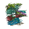
- Structure visualization
| Structure viewer | Molecule: MolmilJmol/JSmol |
|---|
- Downloads & links
-Download
| PDBx/mmCIF format | 6u3y.cif.gz | 350 KB | Display | PDBx/mmCIF format |
|---|---|---|---|---|
| PDB format | pdb6u3y.ent.gz | 285.9 KB | Display | PDB format |
| PDBx/mmJSON format | 6u3y.json.gz | Tree view | PDBx/mmJSON format | |
| Others |  Other downloads Other downloads |
-Validation report
| Arichive directory | https://data.pdbj.org/pub/pdb/validation_reports/u3/6u3yftp://data.pdbj.org/pub/pdb/validation_reports/u3/6u3y | HTTPS FTP |
|---|
-Related structure data
| Related structure data | 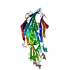 6u2sC 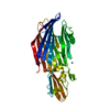 6u33C 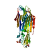 6u3fC 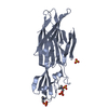 6u3tC  6u49C  6u4pC  3b07S C: citing same article ( S: Starting model for refinement |
|---|---|
| Similar structure data |


-Links
 PDBj
PDBj
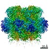
- Assembly
| Deposited unit | 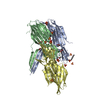
| ||||||||
|---|---|---|---|---|---|---|---|---|---|
| 1 | 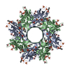
| ||||||||
| 2 | 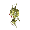
| ||||||||
| Unit cell |
|
-Components
-Protein , 2 types, 3 molecules ABD
| #1: Protein | Mass: 34494.039 Da / Num. of mol.: 1 / Fragment: UNP residues 25-325 Source method: isolated from a genetically manipulated source Source: (gene. exp.) Staphylococcus aureus (bacteria)Gene: lukF-PV, hlgB_1, lukF- PV, LukPV-F, lukPV-F, lukPVF, NCTC6133_01921 Production host: Escherichia coli K-12 (bacteria) / References: UniProt: Q5FBD2 |
|---|---|
| #2: Protein | Mass: 32374.668 Da / Num. of mol.: 2 / Fragment: UNP residues 29-312 Source method: isolated from a genetically manipulated source Source: (gene. exp.) Staphylococcus aureus (bacteria)Gene: lukS-PV, LukPV-S, lukPV-S, lukPVS, EP54_02590, EQ90_09955 Production host: Escherichia coli K-12 (bacteria) / References: UniProt: B1Q017 |
-Non-polymers , 4 types, 376 molecules 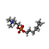


| #3: Chemical |  Mass: 380.523 Da / Num. of mol.: 3 / Source method: obtained synthetically / Formula: C19H43NO4P Mass: 380.523 Da / Num. of mol.: 3 / Source method: obtained synthetically / Formula: C19H43NO4P#4: Chemical | ChemComp-SO4 / Sulfate Mass: 96.063 Da / Num. of mol.: 16 / Source method: obtained synthetically / Formula: SO4 Mass: 96.063 Da / Num. of mol.: 16 / Source method: obtained synthetically / Formula: SO4#5: Chemical | ChemComp-ACT / Acetate Mass: 59.044 Da / Num. of mol.: 8 / Source method: obtained synthetically / Formula: C2H3O2 Mass: 59.044 Da / Num. of mol.: 8 / Source method: obtained synthetically / Formula: C2H3O2#6: Water | ChemComp-HOH / | WaterMass: 18.015 Da / Num. of mol.: 349 / Source method: isolated from a natural source / Formula: H2O |
|---|
-Experimental details
-Experiment
| Experiment | Method: X-RAY DIFFRACTION / Number of used crystals: 1 |
|---|
- Sample preparation
| Crystal | Density Matthews: 3.17 Å3/Da / Density % sol: 61.22 % |
|---|---|
| Crystal grow | Temperature: 298 K / Method: vapor diffusion, hanging drop / pH: 5.4 Details: 6.7 mg/mL LukF-PV, 6.3 mg/mL LukS-PV, 10 mM sodium acetate, pH 5.4, 15 mM Fos-Choline-14, 40 mM beta-OG against reservoir solution of 0.16 M magnesium formate |
-Data collection
| Diffraction | Mean temperature: 100 K / Serial crystal experiment: N |
|---|---|
| Diffraction source | Source: SYNCHROTRON / Site: SSRL / Beamline: BL9-2 / Wavelength: 0.979 Å |
| Detector | Type: DECTRIS PILATUS 6M / Detector: PIXEL / Date: Nov 17, 2018 |
| Radiation | Monochromator: double crystal Si(111) / Protocol: SINGLE WAVELENGTH / Monochromatic (M) / Laue (L): M / Scattering type: x-ray |
| Radiation wavelength | Wavelength: 0.979 Å / Relative weight: 1 |
| Reflection | Resolution: 2.04→38.7 Å / Num. obs: 76882 / % possible obs: 99.9 % / Redundancy: 12.8 % / CC1/2: 0.956 / Rmerge(I) obs: 0.098 / Net I/σ(I): 6.6 |
| Reflection shell | Resolution: 2.04→2.09 Å / Rmerge(I) obs: 0.981 / Mean I/σ(I) obs: 2.1 / Num. unique obs: 3741 / CC1/2: 0.754 |
- Processing
| Software |
| ||||||||||||||||||||||||||||||||||||||||||||||||||||||||||||||||||||||||||||||||||||||||||||||||||||
|---|---|---|---|---|---|---|---|---|---|---|---|---|---|---|---|---|---|---|---|---|---|---|---|---|---|---|---|---|---|---|---|---|---|---|---|---|---|---|---|---|---|---|---|---|---|---|---|---|---|---|---|---|---|---|---|---|---|---|---|---|---|---|---|---|---|---|---|---|---|---|---|---|---|---|---|---|---|---|---|---|---|---|---|---|---|---|---|---|---|---|---|---|---|---|---|---|---|---|---|---|---|
| Refinement | Method to determine structure: MOLECULAR REPLACEMENT Starting model: PDB entry 3B07 Resolution: 2.04→38.69 Å / Cor.coef. Fo:Fc: 0.958 / Cor.coef. Fo:Fc free: 0.943 / SU B: 7.954 / SU ML: 0.105 / Cross valid method: THROUGHOUT / σ(F): 0 / ESU R: 0.152 / ESU R Free: 0.146 / Details: U VALUES : WITH TLS ADDED
| ||||||||||||||||||||||||||||||||||||||||||||||||||||||||||||||||||||||||||||||||||||||||||||||||||||
| Solvent computation | Ion probe radii: 0.8 Å / Shrinkage radii: 0.8 Å / VDW probe radii: 1.2 Å | ||||||||||||||||||||||||||||||||||||||||||||||||||||||||||||||||||||||||||||||||||||||||||||||||||||
| Displacement parameters | Biso max: 129.19 Å2 / Biso mean: 44.123 Å2 / Biso min: 21.94 Å2
| ||||||||||||||||||||||||||||||||||||||||||||||||||||||||||||||||||||||||||||||||||||||||||||||||||||
| Refinement step | Cycle: final / Resolution: 2.04→38.69 Å
| ||||||||||||||||||||||||||||||||||||||||||||||||||||||||||||||||||||||||||||||||||||||||||||||||||||
| Refine LS restraints |
| ||||||||||||||||||||||||||||||||||||||||||||||||||||||||||||||||||||||||||||||||||||||||||||||||||||
| LS refinement shell | Resolution: 2.04→2.092 Å / Rfactor Rfree error: 0
| ||||||||||||||||||||||||||||||||||||||||||||||||||||||||||||||||||||||||||||||||||||||||||||||||||||
| Refinement TLS params. | Method: refined / Refine-ID: X-RAY DIFFRACTION
| ||||||||||||||||||||||||||||||||||||||||||||||||||||||||||||||||||||||||||||||||||||||||||||||||||||
| Refinement TLS group |
|
