 Movie
Movie Controller

[English] 日本語
 Yorodumi
- PDB-6tvz: Structure of a psychrophilic CCA-adding enzyme crystallized in th... -
+ Open data

- Basic information
| Entry | Database: PDB / ID: 6tvz | ||||||||||||
|---|---|---|---|---|---|---|---|---|---|---|---|---|---|
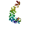
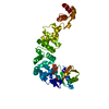
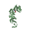
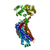
| Title | Structure of a psychrophilic CCA-adding enzyme crystallized in the XtalController device | ||||||||||||
 Components Components | CCA-adding enzyme CCA tRNA nucleotidyltransferase CCA tRNA nucleotidyltransferase | ||||||||||||
 Keywords Keywords | RNA BINDING PROTEIN / tRNA maturation / tRNA nucleotidyltransferase / psychrophilic enzyme | ||||||||||||
| Function / homology |  Function and homology information Function and homology informationRNA 3'-end processing / tRNA processing / nucleotidyltransferase activity / nucleotide binding / RNA binding / metal ion bindingSimilarity search - Function | ||||||||||||
| Biological species |  Planococcus halocryophilus (bacteria) Planococcus halocryophilus (bacteria) | ||||||||||||
| Method | X-RAY DIFFRACTION / SYNCHROTRON / MOLECULAR REPLACEMENT / Resolution: 2.28 Å | ||||||||||||
 Authors Authors | de Wijn, R. / Rollet, K. / Coudray, L. / Hennig, O. / Betat, H. / Moerl, M. / Lorber, B. / Sauter, C. | ||||||||||||
| Funding support |  France, 3items France, 3items
| ||||||||||||
 Citation Citation | Journal: Crystals / Year: 2020 Title: Monitoring the Production of High Diffraction-Quality Crystals of Two Enzymes in Real Time Using In Situ Dynamic Light Scattering Authors: de Wijn, R. / Rollet, K. / Engilberge, S. / McEwen, A.G. / Hennig, O. / Betat, H. / Moerl, M. / Riobe, F. / Maury, O. / Girard, E. / Benas, P. / Lorber, B. / Sauter, C. | ||||||||||||
| History |
|
- Structure visualization
| Structure viewer | Molecule: MolmilJmol/JSmol |
|---|
- Downloads & links
-Download
| PDBx/mmCIF format | 6tvz.cif.gz | 187.7 KB | Display | PDBx/mmCIF format |
|---|---|---|---|---|
| PDB format | pdb6tvz.ent.gz | 131.3 KB | Display | PDB format |
| PDBx/mmJSON format | 6tvz.json.gz | Tree view | PDBx/mmJSON format | |
| Others |  Other downloads Other downloads |
-Validation report
| Arichive directory | https://data.pdbj.org/pub/pdb/validation_reports/tv/6tvzftp://data.pdbj.org/pub/pdb/validation_reports/tv/6tvz | HTTPS FTP |
|---|
-Related structure data
| Related structure data |  6tvyC  6qy6S S: Starting model for refinement C: citing same article ( |
|---|---|
| Similar structure data |


-Links
 PDBj
PDBj- Assembly
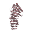
| Deposited unit | 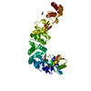
| ||||||||||||
|---|---|---|---|---|---|---|---|---|---|---|---|---|---|
| 1 |
| ||||||||||||
| Unit cell |
|
-Components
| #1: Protein | CCA tRNA nucleotidyltransferase Mass: 48498.266 Da / Num. of mol.: 1 Source method: isolated from a genetically manipulated source Source: (gene. exp.) Planococcus halocryophilus (bacteria) / Gene: BBI08_05760 / Production host: Escherichia coli (E. coli) / References: UniProt: A0A1C7DQ98 | ||||||||
|---|---|---|---|---|---|---|---|---|---|
| #2: Chemical | Phosphate  Mass: 94.971 Da / Num. of mol.: 2 / Source method: isolated from a natural source / Formula: PO4 Mass: 94.971 Da / Num. of mol.: 2 / Source method: isolated from a natural source / Formula: PO4#3: Chemical | Glycerol  Mass: 92.094 Da / Num. of mol.: 3 / Source method: obtained synthetically / Formula: C3H8O3 Mass: 92.094 Da / Num. of mol.: 3 / Source method: obtained synthetically / Formula: C3H8O3#4: Chemical | Acetate  Mass: 59.044 Da / Num. of mol.: 2 / Source method: obtained synthetically / Formula: C2H3O2 Mass: 59.044 Da / Num. of mol.: 2 / Source method: obtained synthetically / Formula: C2H3O2#5: Water | ChemComp-HOH / | Water Mass: 18.015 Da / Num. of mol.: 105 / Source method: isolated from a natural source / Formula: H2O Mass: 18.015 Da / Num. of mol.: 105 / Source method: isolated from a natural source / Formula: H2OHas ligand of interest | N | |
-Experimental details
-Experiment
| Experiment | Method: X-RAY DIFFRACTION / Number of used crystals: 1 |
|---|
- Sample preparation
| Crystal | Density Matthews: 3.74 Å3/Da / Density % sol: 67.4 % / Description: elongated tetragonal bipyramid |
|---|---|
| Crystal grow | Temperature: 298 K / Method: vapor diffusion, sitting drop / pH: 4.5 Details: A 10 micoliter drop of CCA-adding enzyme stock solution at 5 mg/ml in 0.05 M HEPES-NaOH pH 7.5, 0.1 M NaCl was incubated in the XtalController instrument. The crystallization was triggered ...Details: A 10 micoliter drop of CCA-adding enzyme stock solution at 5 mg/ml in 0.05 M HEPES-NaOH pH 7.5, 0.1 M NaCl was incubated in the XtalController instrument. The crystallization was triggered by the gradual addition of crystallant solution (1 M diammonium phosphate, 0.1 M ammonium acetate pH 4.5). |
-Data collection
| Diffraction | Mean temperature: 100 K / Serial crystal experiment: N |
|---|---|
| Diffraction source | Source: SYNCHROTRON / Site: ESRF / Beamline: BM30A / Wavelength: 0.9799 Å |
| Detector | Type: ADSC QUANTUM 315r / Detector: CCD / Date: Apr 23, 2017 |
| Radiation | Protocol: SINGLE WAVELENGTH / Monochromatic (M) / Laue (L): M / Scattering type: x-ray |
| Radiation wavelength | Wavelength: 0.9799 Å / Relative weight: 1 |
| Reflection | Resolution: 2.28→47.2 Å / Num. obs: 34470 / % possible obs: 0.995 % / Redundancy: 6.9 % / Biso Wilson estimate: 44.72 Å2 / CC1/2: 1 / Rpim(I) all: 0.034 / Rrim(I) all: 0.086 / Net I/σ(I): 15.5 |
| Reflection shell | Resolution: 2.28→2.42 Å / Redundancy: 7 % / Num. unique obs: 5385 / CC1/2: 0.76 / Rpim(I) all: 0.448 / Rrim(I) all: 1.26 / % possible all: 98.5 |
- Processing
| Software |
| |||||||||||||||||||||||||||||||||||||||||||||||||||||||||||||||||||||||||||||||||||||||||||
|---|---|---|---|---|---|---|---|---|---|---|---|---|---|---|---|---|---|---|---|---|---|---|---|---|---|---|---|---|---|---|---|---|---|---|---|---|---|---|---|---|---|---|---|---|---|---|---|---|---|---|---|---|---|---|---|---|---|---|---|---|---|---|---|---|---|---|---|---|---|---|---|---|---|---|---|---|---|---|---|---|---|---|---|---|---|---|---|---|---|---|---|---|
| Refinement | Method to determine structure: MOLECULAR REPLACEMENT Starting model: 6QY6 Resolution: 2.28→47.19 Å / SU ML: 0.4031 / Cross valid method: FREE R-VALUE / σ(F): 1.33 / Phase error: 29.9338
| |||||||||||||||||||||||||||||||||||||||||||||||||||||||||||||||||||||||||||||||||||||||||||
| Solvent computation | Shrinkage radii: 0.9 Å / VDW probe radii: 1.11 Å | |||||||||||||||||||||||||||||||||||||||||||||||||||||||||||||||||||||||||||||||||||||||||||
| Displacement parameters | Biso mean: 50.98 Å2 | |||||||||||||||||||||||||||||||||||||||||||||||||||||||||||||||||||||||||||||||||||||||||||
| Refinement step | Cycle: LAST / Resolution: 2.28→47.19 Å
| |||||||||||||||||||||||||||||||||||||||||||||||||||||||||||||||||||||||||||||||||||||||||||
| Refine LS restraints |
| |||||||||||||||||||||||||||||||||||||||||||||||||||||||||||||||||||||||||||||||||||||||||||
| LS refinement shell |
| |||||||||||||||||||||||||||||||||||||||||||||||||||||||||||||||||||||||||||||||||||||||||||
| Refinement TLS params. | Method: refined / Origin x: 40.6403073594 Å / Origin y: 37.0329519812 Å / Origin z: 54.0238436045 Å
| |||||||||||||||||||||||||||||||||||||||||||||||||||||||||||||||||||||||||||||||||||||||||||
| Refinement TLS group | Selection details: all |
