[English] 日本語
Yorodumi
- PDB-6tvq: Structure of native gp41 derived peptide fusion inhibitor -

+
Open data


ID or keywords:

Loading...

-
Basic information

Entry
Database: PDB / ID: 6tvq
TitleStructure of native gp41 derived peptide fusion inhibitor
Components
  • Env polyprotein (Fragment)
  • Envelope glycoprotein gp160
KeywordsVIRAL PROTEIN / Inhibitor / helix bundle / HIV
Function / homology
Function and homology information


virus-mediated perturbation of host defense response => GO:0019049 / : / positive regulation of plasma membrane raft polarization / positive regulation of receptor clustering / positive regulation of establishment of T cell polarity / host cell endosome membrane / clathrin-dependent endocytosis of virus by host cell / membrane => GO:0016020 / viral protein processing / fusion of virus membrane with host plasma membrane ...virus-mediated perturbation of host defense response => GO:0019049 / : / positive regulation of plasma membrane raft polarization / positive regulation of receptor clustering / positive regulation of establishment of T cell polarity / host cell endosome membrane / clathrin-dependent endocytosis of virus by host cell / membrane => GO:0016020 / viral protein processing / fusion of virus membrane with host plasma membrane / fusion of virus membrane with host endosome membrane / viral envelope / virion attachment to host cell / host cell plasma membrane / virion membrane / structural molecule activity / plasma membrane
Similarity search - Function
Envelope glycoprotein Gp160 / Retroviral envelope protein / Retroviral envelope protein GP41-like / Gp120 core superfamily / Envelope glycoprotein GP120 / Human immunodeficiency virus 1, envelope glycoprotein Gp120
Similarity search - Domain/homology
Env polyprotein / Envelope glycoprotein gp160
Similarity search - Component
Biological speciesHuman immunodeficiency virus 1
MethodX-RAY DIFFRACTION / SYNCHROTRON / AB INITIO PHASING / Resolution: 1.45 Å
AuthorsHuhmann, S. / Nyakatura, E.K. / Rohrhofer, A. / Schmidt, B. / Eichler, J. / Moschner, J. / Roth, C.
Funding support Germany, 1items
OrganizationGrant numberCountry
German Research Foundation (DFG) Germany
CitationJournal: Chembiochem / Year: 2021
Title: Systematic Evaluation of Fluorination as Modification for Peptide-Based Fusion Inhibitors against HIV-1 Infection.
Authors: Huhmann, S. / Nyakatura, E.K. / Rohrhofer, A. / Moschner, J. / Schmidt, B. / Eichler, J. / Roth, C. / Koksch, B.
History
DepositionJan 10, 2020Deposition site: PDBE / Processing site: PDBE
Revision 1.0Jan 27, 2021Provider: repository / Type: Initial release
Revision 1.1Feb 16, 2022Group: Database references / Category: citation / citation_author / database_2
Item: _citation.country / _citation.journal_abbrev ..._citation.country / _citation.journal_abbrev / _citation.journal_id_CSD / _citation.journal_id_ISSN / _citation.journal_volume / _citation.page_first / _citation.page_last / _citation.pdbx_database_id_DOI / _citation.pdbx_database_id_PubMed / _citation.title / _citation.year / _database_2.pdbx_DOI / _database_2.pdbx_database_accession
Revision 2.0Jul 19, 2023Group: Atomic model / Data collection ...Atomic model / Data collection / Database references / Derived calculations / Non-polymer description / Polymer sequence / Source and taxonomy / Structure summary
Category: atom_site / atom_site_anisotrop ...atom_site / atom_site_anisotrop / atom_type / chem_comp / entity / entity_poly / entity_poly_seq / pdbx_entity_nonpoly / pdbx_entity_src_syn / pdbx_nonpoly_scheme / pdbx_poly_seq_scheme / pdbx_struct_assembly_gen / pdbx_struct_special_symmetry / struct_asym / struct_conf / struct_conn / struct_ref / struct_ref_seq / struct_ref_seq_dif
Item: _atom_site.B_iso_or_equiv / _atom_site.Cartn_x ..._atom_site.B_iso_or_equiv / _atom_site.Cartn_x / _atom_site.Cartn_y / _atom_site.Cartn_z / _atom_site.auth_asym_id / _atom_site.auth_atom_id / _atom_site.auth_comp_id / _atom_site.auth_seq_id / _atom_site.group_PDB / _atom_site.label_alt_id / _atom_site.label_asym_id / _atom_site.label_atom_id / _atom_site.label_comp_id / _atom_site.label_entity_id / _atom_site.label_seq_id / _atom_site.occupancy / _atom_site.type_symbol / _atom_site_anisotrop.U[1][1] / _atom_site_anisotrop.U[1][2] / _atom_site_anisotrop.U[1][3] / _atom_site_anisotrop.U[2][2] / _atom_site_anisotrop.U[2][3] / _atom_site_anisotrop.U[3][3] / _atom_site_anisotrop.pdbx_auth_asym_id / _atom_site_anisotrop.pdbx_auth_atom_id / _atom_site_anisotrop.pdbx_auth_comp_id / _atom_site_anisotrop.pdbx_auth_seq_id / _atom_site_anisotrop.pdbx_label_alt_id / _atom_site_anisotrop.pdbx_label_asym_id / _atom_site_anisotrop.pdbx_label_atom_id / _atom_site_anisotrop.pdbx_label_comp_id / _atom_site_anisotrop.pdbx_label_seq_id / _atom_site_anisotrop.type_symbol / _atom_type.pdbx_N_electrons / _atom_type.pdbx_scat_Z / _chem_comp.formula / _chem_comp.formula_weight / _chem_comp.id / _chem_comp.name / _entity_poly.nstd_monomer / _entity_poly.pdbx_seq_one_letter_code / _entity_poly.pdbx_seq_one_letter_code_can / _pdbx_entity_src_syn.pdbx_end_seq_num / _pdbx_struct_assembly_gen.asym_id_list / _pdbx_struct_special_symmetry.label_asym_id / _struct_conf.beg_auth_seq_id / _struct_conf.beg_label_seq_id / _struct_conf.end_auth_seq_id / _struct_conf.end_label_seq_id / _struct_conn.pdbx_leaving_atom_flag / _struct_conn.ptnr1_auth_comp_id / _struct_conn.ptnr1_auth_seq_id / _struct_conn.ptnr1_label_atom_id / _struct_conn.ptnr1_label_comp_id / _struct_conn.ptnr1_label_seq_id / _struct_conn.ptnr2_auth_comp_id / _struct_conn.ptnr2_auth_seq_id / _struct_conn.ptnr2_label_asym_id / _struct_conn.ptnr2_label_atom_id / _struct_conn.ptnr2_label_comp_id / _struct_conn.ptnr2_label_seq_id / _struct_ref.db_code / _struct_ref.pdbx_align_begin / _struct_ref.pdbx_db_accession / _struct_ref_seq.db_align_beg / _struct_ref_seq.db_align_end / _struct_ref_seq.pdbx_auth_seq_align_beg / _struct_ref_seq.pdbx_auth_seq_align_end / _struct_ref_seq.pdbx_db_accession / _struct_ref_seq.seq_align_beg / _struct_ref_seq.seq_align_end

-
Structure visualization

Structure viewerMolecule:
MolmilJmol/JSmol

Downloads & links

-
Assembly

Deposited unit
AaA: Env polyprotein (Fragment)
BBB: Envelope glycoprotein gp160


Theoretical massNumber of molelcules
Total (without water)8,4292
Polymers8,4292
Non-polymers00
Water95553
1
AaA: Env polyprotein (Fragment)
BBB: Envelope glycoprotein gp160

AaA: Env polyprotein (Fragment)
BBB: Envelope glycoprotein gp160

AaA: Env polyprotein (Fragment)
BBB: Envelope glycoprotein gp160


Theoretical massNumber of molelcules
Total (without water)25,2866
Polymers25,2866
Non-polymers00
Water1086
TypeNameSymmetry operationNumber
identity operation1_555x,y,z1
crystal symmetry operation2_455-y-1,x-y,z1
crystal symmetry operation3_445-x+y-1,-x-1,z1
Buried area10620 Å2
ΔGint-85 kcal/mol
Surface area10540 Å2
MethodPISA
Unit cell
Length a, b, c (Å)44.875, 44.875, 209.645
Angle α, β, γ (deg.)90.000, 90.000, 120.000
Int Tables number155
Space group name H-MH32
Components on special symmetry positions
IDModelComponents
11AaA-705-

HOH

21AaA-716-

HOH

-
Components

#1: Protein/peptide Env polyprotein (Fragment)


Mass: 4515.291 Da / Num. of mol.: 1 / Source method: obtained synthetically / Source: (synth.) Human immunodeficiency virus 1 / References: UniProt: C7F3P9
#2: Protein/peptide Envelope glycoprotein gp160 / Env polyprotein


Mass: 3913.264 Da / Num. of mol.: 1 / Source method: obtained synthetically / Source: (synth.) Human immunodeficiency virus 1 / References: UniProt: Q6TAQ1
#3: Water ChemComp-HOH / water / Water


Mass: 18.015 Da / Num. of mol.: 53 / Source method: isolated from a natural source / Formula: H2O
Has ligand of interestN

-
Experimental details

-
Experiment

ExperimentMethod: X-RAY DIFFRACTION / Number of used crystals: 1

-
Sample preparation

CrystalDensity Matthews: 2.42 Å3/Da / Density % sol: 49.11 %
Crystal growTemperature: 292.15 K / Method: vapor diffusion, sitting drop / pH: 5.5 / Details: Ammonium sulfate, PEG 3350, Bis-Tris

-
Data collection

DiffractionMean temperature: 100 K / Serial crystal experiment: N
Diffraction sourceSource: SYNCHROTRON / Site: BESSY / Beamline: 14.1 / Wavelength: 0.9184 Å
DetectorType: DECTRIS PILATUS3 S 6M / Detector: PIXEL / Date: Nov 6, 2019
RadiationProtocol: SINGLE WAVELENGTH / Monochromatic (M) / Laue (L): M / Scattering type: x-ray
Radiation wavelengthWavelength: 0.9184 Å / Relative weight: 1
ReflectionResolution: 1.45→38.241 Å / Num. obs: 14982 / % possible obs: 100 % / Redundancy: 11.1 % / CC1/2: 0.997 / Rmerge(I) obs: 0.152 / Rpim(I) all: 0.048 / Net I/σ(I): 8
Reflection shellResolution: 1.45→1.48 Å / Mean I/σ(I) obs: 0.9 / Num. unique obs: 742 / CC1/2: 0.669

-
Processing

Software
NameVersionClassification
REFMAC5.8.0258refinement
XDSdata reduction
Aimlessdata scaling
ACORNphasing
RefinementMethod to determine structure: AB INITIO PHASING / Resolution: 1.45→38.24 Å / Cor.coef. Fo:Fc: 0.966 / Cor.coef. Fo:Fc free: 0.95 / SU B: 4.661 / SU ML: 0.073 / Cross valid method: FREE R-VALUE / ESU R: 0.073 / ESU R Free: 0.074
Details: Hydrogens have been added in their riding positions
RfactorNum. reflection% reflection
Rfree0.2426 710 4.74 %
Rwork0.1879 --
all0.19 --
obs-14980 99.953 %
Solvent computationIon probe radii: 0.8 Å / Shrinkage radii: 0.8 Å / VDW probe radii: 1.2 Å
Displacement parametersBiso mean: 27.725 Å2
Baniso -1Baniso -2Baniso -3
1-1.856 Å20.928 Å2-0 Å2
2--1.856 Å2-0 Å2
3----6.02 Å2
Refinement stepCycle: LAST / Resolution: 1.45→38.24 Å
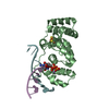
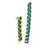
ProteinNucleic acidLigandSolventTotal
Num. atoms535 0 4 53 592
Refine LS restraints
Refine-IDTypeDev idealDev ideal targetNumber
X-RAY DIFFRACTIONr_bond_refined_d0.0130.013563
X-RAY DIFFRACTIONr_bond_other_d0.0010.017521
X-RAY DIFFRACTIONr_angle_refined_deg1.711.633765
X-RAY DIFFRACTIONr_angle_other_deg1.5851.5711206
X-RAY DIFFRACTIONr_dihedral_angle_1_deg5.355565
X-RAY DIFFRACTIONr_dihedral_angle_2_deg26.12821.48127
X-RAY DIFFRACTIONr_dihedral_angle_3_deg13.9861593
X-RAY DIFFRACTIONr_dihedral_angle_4_deg15.095154
X-RAY DIFFRACTIONr_chiral_restr0.0860.274
X-RAY DIFFRACTIONr_gen_planes_refined0.0090.02628
X-RAY DIFFRACTIONr_gen_planes_other0.0030.02117
X-RAY DIFFRACTIONr_nbd_refined0.2180.2134
X-RAY DIFFRACTIONr_symmetry_nbd_other0.1740.2421
X-RAY DIFFRACTIONr_nbtor_refined0.1740.2272
X-RAY DIFFRACTIONr_symmetry_nbtor_other0.0860.2246
X-RAY DIFFRACTIONr_xyhbond_nbd_refined0.3110.225
X-RAY DIFFRACTIONr_symmetry_nbd_refined0.2530.215
X-RAY DIFFRACTIONr_nbd_other0.1830.259
X-RAY DIFFRACTIONr_symmetry_xyhbond_nbd_refined0.1330.25
X-RAY DIFFRACTIONr_mcbond_it4.1382.61260
X-RAY DIFFRACTIONr_mcbond_other4.1322.619261
X-RAY DIFFRACTIONr_mcangle_it5.0233.92325
X-RAY DIFFRACTIONr_mcangle_other5.0163.931326
X-RAY DIFFRACTIONr_scbond_it5.3293.167303
X-RAY DIFFRACTIONr_scbond_other5.3033.154302
X-RAY DIFFRACTIONr_scangle_it6.3624.579440
X-RAY DIFFRACTIONr_scangle_other6.3554.59441
X-RAY DIFFRACTIONr_lrange_it5.61632.464667
X-RAY DIFFRACTIONr_lrange_other5.24732.222657
X-RAY DIFFRACTIONr_rigid_bond_restr4.10931082
LS refinement shell
Resolution (Å)Rfactor RfreeNum. reflection RfreeRfactor RworkNum. reflection RworkRefine-ID% reflection obs (%)
1.45-1.4880.413460.3651042X-RAY DIFFRACTION100
1.488-1.5290.333650.309990X-RAY DIFFRACTION100
1.529-1.5730.312510.267963X-RAY DIFFRACTION99.9015
1.573-1.6210.316540.256963X-RAY DIFFRACTION100
1.621-1.6740.295470.227920X-RAY DIFFRACTION100
1.674-1.7330.238460.194902X-RAY DIFFRACTION100
1.733-1.7980.234380.175869X-RAY DIFFRACTION100
1.798-1.8720.204370.14844X-RAY DIFFRACTION100
1.872-1.9550.263460.165805X-RAY DIFFRACTION100
1.955-2.050.204310.164774X-RAY DIFFRACTION99.8759
2.05-2.1610.194260.135743X-RAY DIFFRACTION100
2.161-2.2920.2250.133705X-RAY DIFFRACTION99.8632
2.292-2.450.205280.149659X-RAY DIFFRACTION100
2.45-2.6460.213370.153623X-RAY DIFFRACTION100
2.646-2.8980.207350.171558X-RAY DIFFRACTION100
2.898-3.240.302240.224532X-RAY DIFFRACTION100
3.24-3.740.274300.19456X-RAY DIFFRACTION100
3.74-4.5770.203180.152400X-RAY DIFFRACTION99.7613
4.577-6.4590.178150.191323X-RAY DIFFRACTION100
6.459-100.266110.247199X-RAY DIFFRACTION100

+
About Yorodumi

-
News

-
Feb 9, 2022. New format data for meta-information of EMDB entries

New format data for meta-information of EMDB entries

  • Version 3 of the EMDB header file is now the official format.
  • The previous official version 1.9 will be removed from the archive.

Related info.:EMDB header

External links:wwPDB to switch to version 3 of the EMDB data model

-
Aug 12, 2020. Covid-19 info

Covid-19 info

URL: https://pdbj.org/emnavi/covid19.php

New page: Covid-19 featured information page in EM Navigator.

Related info.:Covid-19 info / Mar 5, 2020. Novel coronavirus structure data

+
Mar 5, 2020. Novel coronavirus structure data

Novel coronavirus structure data

Related info.:Yorodumi Speices / Aug 12, 2020. Covid-19 info

External links:COVID-19 featured content - PDBj / Molecule of the Month (242):Coronavirus Proteases

+
Jan 31, 2019. EMDB accession codes are about to change! (news from PDBe EMDB page)

EMDB accession codes are about to change! (news from PDBe EMDB page)

  • The allocation of 4 digits for EMDB accession codes will soon come to an end. Whilst these codes will remain in use, new EMDB accession codes will include an additional digit and will expand incrementally as the available range of codes is exhausted. The current 4-digit format prefixed with “EMD-” (i.e. EMD-XXXX) will advance to a 5-digit format (i.e. EMD-XXXXX), and so on. It is currently estimated that the 4-digit codes will be depleted around Spring 2019, at which point the 5-digit format will come into force.
  • The EM Navigator/Yorodumi systems omit the EMD- prefix.

Related info.:Q: What is EMD? / ID/Accession-code notation in Yorodumi/EM Navigator

External links:EMDB Accession Codes are Changing Soon! / Contact to PDBj

+
Jul 12, 2017. Major update of PDB

Major update of PDB

  • wwPDB released updated PDB data conforming to the new PDBx/mmCIF dictionary.
  • This is a major update changing the version number from 4 to 5, and with Remediation, in which all the entries are updated.
  • In this update, many items about electron microscopy experimental information are reorganized (e.g. em_software).
  • Now, EM Navigator and Yorodumi are based on the updated data.

External links:wwPDB Remediation / Enriched Model Files Conforming to OneDep Data Standards Now Available in the PDB FTP Archive

-
Yorodumi

Thousand views of thousand structures

  • Yorodumi is a browser for structure data from EMDB, PDB, SASBDB, etc.
  • This page is also the successor to EM Navigator detail page, and also detail information page/front-end page for Omokage search.
  • The word "yorodu" (or yorozu) is an old Japanese word meaning "ten thousand". "mi" (miru) is to see.

Related info.:EMDB / PDB / SASBDB / Comparison of 3 databanks / Yorodumi Search / Aug 31, 2016. New EM Navigator & Yorodumi / Yorodumi Papers / Jmol/JSmol / Function and homology information / Changes in new EM Navigator and Yorodumi

Read more