 Movie
Movie Controller

[English] 日本語
 Yorodumi
- PDB-6tuo: Crystal structure of Archaeoglobus fulgidus Argonaute protein wit... -
+ Open data

- Basic information
| Entry | Database: PDB / ID: 6tuo | ||||||
|---|---|---|---|---|---|---|---|
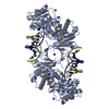
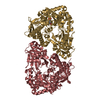
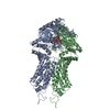
| Title | Crystal structure of Archaeoglobus fulgidus Argonaute protein with cognate DNA oligoduplex 5'-pATTGTACGTACAAT | ||||||
 Components Components |
| ||||||
 Keywords Keywords |  DNA BINDING PROTEIN / ARGONAUTE / PIWI DOMAIN / PROTEIN-DNA COMPLEX DNA BINDING PROTEIN / ARGONAUTE / PIWI DOMAIN / PROTEIN-DNA COMPLEX | ||||||
| Function / homology |  Function and homology information Function and homology information | ||||||
| Biological species |   Archaeoglobus fulgidus (archaea) Archaeoglobus fulgidus (archaea) Escherichia coli (E. coli) Escherichia coli (E. coli) | ||||||
| Method | X-RAY DIFFRACTION / SYNCHROTRON / MOLECULAR REPLACEMENT / molecular replacement / Resolution: 1.8 Å | ||||||
 Authors Authors | Grazulis, S. / Zaremba, M. | ||||||
 Citation Citation | Journal: To be published Title: Crystal structure of Archaeoglobus fulgidus Argonaute protein with cognate DNA oligoduplex 5'-pATTGTACGTACAAT Authors: Golovinas, E. / Manakova, E. / Sasnauskas, G. / Zaremba, M. | ||||||
| History |
|
- Structure visualization
| Structure viewer | Molecule: MolmilJmol/JSmol |
|---|
- Downloads & links
-Download
| PDBx/mmCIF format | 6tuo.cif.gz | 132.3 KB | Display | PDBx/mmCIF format |
|---|---|---|---|---|
| PDB format | pdb6tuo.ent.gz | 95.8 KB | Display | PDB format |
| PDBx/mmJSON format | 6tuo.json.gz | Tree view | PDBx/mmJSON format | |
| Others |  Other downloads Other downloads |
-Validation report
| Arichive directory | https://data.pdbj.org/pub/pdb/validation_reports/tu/6tuoftp://data.pdbj.org/pub/pdb/validation_reports/tu/6tuo | HTTPS FTP |
|---|
-Related structure data
| Related structure data | 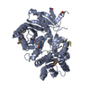 1ytuS S: Starting model for refinement |
|---|---|
| Similar structure data |


-Links
 PDBj
PDBj


- Assembly
| Deposited unit | 
| |||||||||
|---|---|---|---|---|---|---|---|---|---|---|
| 1 | 
| |||||||||
| Unit cell |
| |||||||||
| Components on special symmetry positions |
|
-Components
-Protein / DNA chain , 2 types, 3 molecules ARS
| #1: Protein | / AfAgo / AfPiwi / PIWI/MID domain protein Mass: 50921.121 Da / Num. of mol.: 1 / Fragment: Arhaeoglobus fulgidus Argonaute protein Source method: isolated from a genetically manipulated source Source: (gene. exp.) Archaeoglobus fulgidus (strain ATCC 49558 / VC-16 / DSM 4304 / JCM 9628 / NBRC 100126) (archaea)Strain: DSM 4304 / Gene: AF_1318 / Plasmid: pETDuet / Production host: Escherichia coli BL21(DE3) (bacteria) / References: UniProt: O28951 |
|---|---|
| #2: DNA chain | Mass: 4278.815 Da / Num. of mol.: 2 / Fragment: oligodeoxyribonucleotide / Source method: obtained synthetically / Source: (synth.) Escherichia coli (E. coli) |
-Non-polymers , 6 types, 360 molecules 


| #3: Chemical | ChemComp-MG /  Mass: 24.305 Da / Num. of mol.: 1 / Fragment: oligodeoxyribonucleotide / Source method: obtained synthetically / Formula: Mg Mass: 24.305 Da / Num. of mol.: 1 / Fragment: oligodeoxyribonucleotide / Source method: obtained synthetically / Formula: Mg | ||||||
|---|---|---|---|---|---|---|---|
| #4: Chemical | ChemComp-CL / Chloride Mass: 35.453 Da / Num. of mol.: 1 / Source method: obtained synthetically / Formula: Cl Mass: 35.453 Da / Num. of mol.: 1 / Source method: obtained synthetically / Formula: Cl | ||||||
| #5: Chemical | 2-Mercaptoethanol Mass: 78.133 Da / Num. of mol.: 3 / Source method: obtained synthetically / Formula: C2H6OS Mass: 78.133 Da / Num. of mol.: 3 / Source method: obtained synthetically / Formula: C2H6OS#6: Chemical | ChemComp-PEG / | Diethylene glycol Mass: 106.120 Da / Num. of mol.: 1 / Source method: obtained synthetically / Formula: C4H10O3 Mass: 106.120 Da / Num. of mol.: 1 / Source method: obtained synthetically / Formula: C4H10O3#7: Chemical | ChemComp-GOL / Glycerol Mass: 92.094 Da / Num. of mol.: 4 / Source method: obtained synthetically / Formula: C3H8O3 Mass: 92.094 Da / Num. of mol.: 4 / Source method: obtained synthetically / Formula: C3H8O3#8: Water | ChemComp-HOH / | WaterMass: 18.015 Da / Num. of mol.: 350 / Source method: isolated from a natural source / Formula: H2O |
-Details
| Has ligand of interest | N |
|---|
-Experimental details
-Experiment
| Experiment | Method: X-RAY DIFFRACTION / Number of used crystals: 1 |
|---|
- Sample preparation
| Crystal | Density Matthews: 2.4 Å3/Da / Density % sol: 48.66 % |
|---|---|
| Crystal grow | Temperature: 280 K / Method: vapor diffusion, sitting drop / pH: 5.5 Details: Na-Cacodylate pH 5.5 0.05 M, KCl_0.2 M, MgCl2 0.01 M, PEG4000 5% (w/v), glycerol 5% (v/v) |
-Data collection
| Diffraction | Mean temperature: 100 K / Serial crystal experiment: N | ||||||||||||||||||||||||||||||
|---|---|---|---|---|---|---|---|---|---|---|---|---|---|---|---|---|---|---|---|---|---|---|---|---|---|---|---|---|---|---|---|
| Diffraction source | Source: SYNCHROTRON / Site: PETRA III, EMBL c/o DESY  / Beamline: P13 (MX1) / Wavelength: 0.9797 Å / Beamline: P13 (MX1) / Wavelength: 0.9797 Å | ||||||||||||||||||||||||||||||
| Detector | Type: DECTRIS PILATUS3 6M / Detector: PIXEL / Date: Nov 23, 2018 / Details: silica substrate + Rh coating | ||||||||||||||||||||||||||||||
| Radiation | Monochromator: Oxford-FMB, UK; Si(111) or Si(311); both crystals LN2 cooled, fixed exit Protocol: SINGLE WAVELENGTH / Monochromatic (M) / Laue (L): M / Scattering type: x-ray | ||||||||||||||||||||||||||||||
| Radiation wavelength | Wavelength: 0.9797 Å / Relative weight: 1 | ||||||||||||||||||||||||||||||
| Reflection | Resolution: 1.8→99.63 Å / Num. all: 349485 / Num. obs: 53664 / % possible obs: 99.9 % / Redundancy: 6.5 % / Biso Wilson estimate: 29.43 Å2 / CC1/2: 0.999 / Rmerge(I) obs: 0.044 / Rpim(I) all: 0.019 / Rrim(I) all: 0.048 / Net I/σ(I): 20.8 | ||||||||||||||||||||||||||||||
| Reflection shell | Diffraction-ID: 1
|
-Phasing
| Phasing | Method: molecular replacement | |||||||||
|---|---|---|---|---|---|---|---|---|---|---|
| Phasing MR | R rigid body: 0.461
|
- Processing
| Software |
| ||||||||||||||||||||||||||||||||||||||||||||||||||||||||||||||||||||||||||||||||||||||||||||||||||||||||||||||||||||||||||||||||||||||||||||||||||||||||||||||||||||||||||||||||||||||||||
|---|---|---|---|---|---|---|---|---|---|---|---|---|---|---|---|---|---|---|---|---|---|---|---|---|---|---|---|---|---|---|---|---|---|---|---|---|---|---|---|---|---|---|---|---|---|---|---|---|---|---|---|---|---|---|---|---|---|---|---|---|---|---|---|---|---|---|---|---|---|---|---|---|---|---|---|---|---|---|---|---|---|---|---|---|---|---|---|---|---|---|---|---|---|---|---|---|---|---|---|---|---|---|---|---|---|---|---|---|---|---|---|---|---|---|---|---|---|---|---|---|---|---|---|---|---|---|---|---|---|---|---|---|---|---|---|---|---|---|---|---|---|---|---|---|---|---|---|---|---|---|---|---|---|---|---|---|---|---|---|---|---|---|---|---|---|---|---|---|---|---|---|---|---|---|---|---|---|---|---|---|---|---|---|---|---|---|---|
| Refinement | Method to determine structure: MOLECULAR REPLACEMENT Starting model: pdbid 1ytu Resolution: 1.8→54.901 Å / SU ML: 0.21 / Cross valid method: THROUGHOUT / σ(F): 0.89 / Phase error: 27.38
| ||||||||||||||||||||||||||||||||||||||||||||||||||||||||||||||||||||||||||||||||||||||||||||||||||||||||||||||||||||||||||||||||||||||||||||||||||||||||||||||||||||||||||||||||||||||||||
| Solvent computation | Shrinkage radii: 0.9 Å / VDW probe radii: 1.11 Å | ||||||||||||||||||||||||||||||||||||||||||||||||||||||||||||||||||||||||||||||||||||||||||||||||||||||||||||||||||||||||||||||||||||||||||||||||||||||||||||||||||||||||||||||||||||||||||
| Displacement parameters | Biso max: 169.13 Å2 / Biso mean: 48.285 Å2 / Biso min: 15.24 Å2 | ||||||||||||||||||||||||||||||||||||||||||||||||||||||||||||||||||||||||||||||||||||||||||||||||||||||||||||||||||||||||||||||||||||||||||||||||||||||||||||||||||||||||||||||||||||||||||
| Refinement step | Cycle: final / Resolution: 1.8→54.901 Å
| ||||||||||||||||||||||||||||||||||||||||||||||||||||||||||||||||||||||||||||||||||||||||||||||||||||||||||||||||||||||||||||||||||||||||||||||||||||||||||||||||||||||||||||||||||||||||||
| LS refinement shell | Refine-ID: X-RAY DIFFRACTION / Rfactor Rfree error: 0
|
