 Movie
Movie Controller


+ Open data

- Basic information
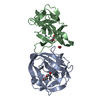
| Entry | Database: PDB / ID: 6tmm | ||||||
|---|---|---|---|---|---|---|---|
| Title | BIL2 domain from T.thermophila BUBL1 locus (C1A-N143A) | ||||||
 Components Components | (NAD(P)(+)--arginine ADP- ...) x 2 | ||||||
 Keywords Keywords |  SPLICING / Intein / PTM / ubiquitin SPLICING / Intein / PTM / ubiquitin | ||||||
| Function / homology |  Function and homology information Function and homology informationNAD+-protein-arginine ADP-ribosyltransferase / NAD+-protein-arginine ADP-ribosyltransferase activity / intein-mediated protein splicing / nucleotidyltransferase activitySimilarity search - Function | ||||||
| Biological species |  Tetrahymena thermophila SB210 (eukaryote) Tetrahymena thermophila SB210 (eukaryote) | ||||||
| Method | X-RAY DIFFRACTION / SYNCHROTRON / SAD / Resolution: 2.398 Å | ||||||
 Authors Authors | Chiarini, V. / Ilari, A. | ||||||
| Funding support |  Italy, 1items Italy, 1items
| ||||||
 Citation Citation | Journal: Biochim Biophys Acta Gen Subj / Year: 2021 Title: Structural basis of ubiquitination mediated by protein splicing in early Eukarya. Authors: Chiarini, V. / Fiorillo, A. / Camerini, S. / Crescenzi, M. / Nakamura, S. / Battista, T. / Guidoni, L. / Colotti, G. / Ilari, A. #1: Journal: Biochim.Biophys.Acta / Year: 2021Title: Structural basis of ubiquitination mediated by protein splicing in early Eukarya Authors: Chiarini, V. / Fiorillo, A. / Camerini, S. / Crescenzi, M. / Nakamura, S. / Battista, T. / Guidoni, L. / Colotti, G. / Ilari, A. | ||||||
| History |
|
- Structure visualization
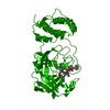
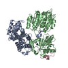
| Structure viewer | Molecule: MolmilJmol/JSmol |
|---|
- Downloads & links
-Download
| PDBx/mmCIF format | 6tmm.cif.gz | 140.4 KB | Display | PDBx/mmCIF format |
|---|---|---|---|---|
| PDB format | pdb6tmm.ent.gz | Display | PDB format | |
| PDBx/mmJSON format | 6tmm.json.gz | Tree view | PDBx/mmJSON format | |
| Others |  Other downloads Other downloads |
-Validation report
| Arichive directory | https://data.pdbj.org/pub/pdb/validation_reports/tm/6tmmftp://data.pdbj.org/pub/pdb/validation_reports/tm/6tmm | HTTPS FTP |
|---|
-Related structure data


-Links
 PDBj
PDBj


- Assembly
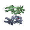
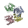
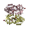
| Deposited unit | 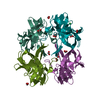
| ||||||||
|---|---|---|---|---|---|---|---|---|---|
| 1 |
| ||||||||
| Unit cell |
| ||||||||
| Components on special symmetry positions |
| ||||||||
| Noncrystallographic symmetry (NCS) | NCS domain: (Details: Chains AAA BBB CCC DDD) |
-Components
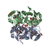
-NAD(P)(+)--arginine ADP- ... , 2 types, 4 molecules AAACCCBBBDDD
| #1: Protein | Mass: 17842.262 Da / Num. of mol.: 2 Source method: isolated from a genetically manipulated source Source: (gene. exp.) Tetrahymena thermophila SB210 (eukaryote)Gene: TTHERM_00085190 / Production host:  Escherichia coli BL21(DE3) (bacteria) Escherichia coli BL21(DE3) (bacteria)References: UniProt: Q236S9, NAD+-protein-arginine ADP-ribosyltransferase #2: Protein | Mass: 17729.104 Da / Num. of mol.: 2 Source method: isolated from a genetically manipulated source Source: (gene. exp.) Tetrahymena thermophila SB210 (eukaryote)Gene: TTHERM_00085190 / Production host: Escherichia coli BL21(DE3) (bacteria)References: UniProt: Q236S9, NAD+-protein-arginine ADP-ribosyltransferase |
|---|
-Non-polymers , 5 types, 140 molecules 


| #3: Chemical | ChemComp-FMT / Formic acid Mass: 46.025 Da / Num. of mol.: 11 / Source method: obtained synthetically / Formula: CH2O2 Mass: 46.025 Da / Num. of mol.: 11 / Source method: obtained synthetically / Formula: CH2O2#4: Chemical | ChemComp-PEG / | Diethylene glycol Mass: 106.120 Da / Num. of mol.: 1 / Source method: obtained synthetically / Formula: C4H10O3 Mass: 106.120 Da / Num. of mol.: 1 / Source method: obtained synthetically / Formula: C4H10O3#5: Chemical | ChemComp-HG / Mercury (element) Mass: 200.590 Da / Num. of mol.: 4 / Source method: obtained synthetically / Formula: Hg Mass: 200.590 Da / Num. of mol.: 4 / Source method: obtained synthetically / Formula: Hg#6: Chemical | ChemComp-CA / |  Mass: 40.078 Da / Num. of mol.: 1 / Source method: obtained synthetically / Formula: Ca Mass: 40.078 Da / Num. of mol.: 1 / Source method: obtained synthetically / Formula: Ca#7: Water | ChemComp-HOH / | WaterMass: 18.015 Da / Num. of mol.: 123 / Source method: isolated from a natural source / Formula: H2O |
|---|
-Details
| Has ligand of interest | N |
|---|
-Experimental details
-Experiment
| Experiment | Method: X-RAY DIFFRACTION / Number of used crystals: 1 |
|---|
- Sample preparation
| Crystal | Density Matthews: 2.35 Å3/Da / Density % sol: 47.63 % |
|---|---|
| Crystal grow | Temperature: 293 K / Method: vapor diffusion, hanging drop / pH: 8.6 Details: Mg formate 0.1 M, PEG3350 19%, Tris 50 mM, NaCl 250 mM. |
-Data collection
| Diffraction | Mean temperature: 100 K / Serial crystal experiment: N |
|---|---|
| Diffraction source | Source: SYNCHROTRON / Site: ELETTRA / Beamline: 11.2C / Wavelength: 1 Å |
| Detector | Type: DECTRIS PILATUS3 S 6M / Detector: PIXEL / Date: Nov 14, 2019 |
| Radiation | Protocol: SINGLE WAVELENGTH / Monochromatic (M) / Laue (L): M / Scattering type: x-ray |
| Radiation wavelength | Wavelength: 1 Å / Relative weight: 1 |
| Reflection | Resolution: 2.398→69.602 Å / Num. obs: 26317 / % possible obs: 98.9 % / Redundancy: 3.36 % / Biso Wilson estimate: 58.51 Å2 / CC1/2: 0.998 / Rmerge(I) obs: 0.06 / Rrim(I) all: 0.071 / Net I/σ(I): 12.25 |
| Reflection shell | Resolution: 2.398→2.54 Å / Redundancy: 3.37 % / Rmerge(I) obs: 0.593 / Mean I/σ(I) obs: 1.78 / Num. unique obs: 8236 / CC1/2: 0.733 / Rrim(I) all: 0.704 / % possible all: 98.9 |
- Processing
| Software |
| ||||||||||||||||||||||||||||||||||||||||||||||||||||||||||||||||||||||||||||||||||||||||||||||||||||||||||||||||||||||||||||||||||||||||||||||||||||||||||||||||
|---|---|---|---|---|---|---|---|---|---|---|---|---|---|---|---|---|---|---|---|---|---|---|---|---|---|---|---|---|---|---|---|---|---|---|---|---|---|---|---|---|---|---|---|---|---|---|---|---|---|---|---|---|---|---|---|---|---|---|---|---|---|---|---|---|---|---|---|---|---|---|---|---|---|---|---|---|---|---|---|---|---|---|---|---|---|---|---|---|---|---|---|---|---|---|---|---|---|---|---|---|---|---|---|---|---|---|---|---|---|---|---|---|---|---|---|---|---|---|---|---|---|---|---|---|---|---|---|---|---|---|---|---|---|---|---|---|---|---|---|---|---|---|---|---|---|---|---|---|---|---|---|---|---|---|---|---|---|---|---|---|---|
| Refinement | Method to determine structure: SAD / Resolution: 2.398→69.602 Å / Cor.coef. Fo:Fc: 0.96 / Cor.coef. Fo:Fc free: 0.925 / SU B: 10.873 / SU ML: 0.241 / Cross valid method: THROUGHOUT / ESU R: 0.55 / ESU R Free: 0.298 Details: Hydrogens have been added in their riding positions
| ||||||||||||||||||||||||||||||||||||||||||||||||||||||||||||||||||||||||||||||||||||||||||||||||||||||||||||||||||||||||||||||||||||||||||||||||||||||||||||||||
| Solvent computation | Ion probe radii: 0.8 Å / Shrinkage radii: 0.8 Å / VDW probe radii: 1.2 Å | ||||||||||||||||||||||||||||||||||||||||||||||||||||||||||||||||||||||||||||||||||||||||||||||||||||||||||||||||||||||||||||||||||||||||||||||||||||||||||||||||
| Displacement parameters | Biso mean: 56.817 Å2
| ||||||||||||||||||||||||||||||||||||||||||||||||||||||||||||||||||||||||||||||||||||||||||||||||||||||||||||||||||||||||||||||||||||||||||||||||||||||||||||||||
| Refinement step | Cycle: LAST / Resolution: 2.398→69.602 Å
| ||||||||||||||||||||||||||||||||||||||||||||||||||||||||||||||||||||||||||||||||||||||||||||||||||||||||||||||||||||||||||||||||||||||||||||||||||||||||||||||||
| Refine LS restraints |
| ||||||||||||||||||||||||||||||||||||||||||||||||||||||||||||||||||||||||||||||||||||||||||||||||||||||||||||||||||||||||||||||||||||||||||||||||||||||||||||||||
| LS refinement shell |
|
