 Movie
Movie Controller


+ Open data

- Basic information
| Entry | Database: PDB / ID: 6tmd | ||||||||||||
|---|---|---|---|---|---|---|---|---|---|---|---|---|---|
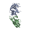
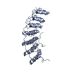
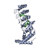
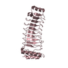
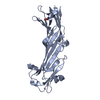
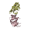
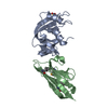
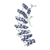
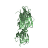
| Title | Crystal structure of KANK2 ankyrin repeats mutant (A670V) | ||||||||||||
 Components Components | KN motif and ankyrin repeat domain-containing protein 2 | ||||||||||||
 Keywords Keywords |  CELL ADHESION / Protein binding / ANKYRIN REPEAT DOMAIN-CONTAINING PROTEIN 25 / MATRIX-REMODELING-ASSOCIATED PROTEIN 3 CELL ADHESION / Protein binding / ANKYRIN REPEAT DOMAIN-CONTAINING PROTEIN 25 / MATRIX-REMODELING-ASSOCIATED PROTEIN 3 | ||||||||||||
| Function / homology |  Function and homology information Function and homology informationnegative regulation of vitamin D receptor signaling pathway / kidney epithelium development / podocyte cell migration / negative regulation of intracellular estrogen receptor signaling pathway / negative regulation of actin filament polymerization / regulation of Rho protein signal transduction / negative regulation of programmed cell death / negative regulation of G1/S transition of mitotic cell cycle / negative regulation of cell population proliferation / apoptotic process ...negative regulation of vitamin D receptor signaling pathway / kidney epithelium development / podocyte cell migration / negative regulation of intracellular estrogen receptor signaling pathway / negative regulation of actin filament polymerization / regulation of Rho protein signal transduction / negative regulation of programmed cell death / negative regulation of G1/S transition of mitotic cell cycle / negative regulation of cell population proliferation / apoptotic process / negative regulation of transcription by RNA polymerase II / mitochondrion / cytoplasmSimilarity search - Function | ||||||||||||
| Biological species |  Homo sapiens (human) Homo sapiens (human) | ||||||||||||
| Method | X-RAY DIFFRACTION / SYNCHROTRON / MOLECULAR REPLACEMENT / Resolution: 1.5 Å | ||||||||||||
 Authors Authors | Khan, R. / Singh, A.K. / Goult, B.T. | ||||||||||||
| Funding support |  United Kingdom, 3items United Kingdom, 3items
| ||||||||||||
 Citation Citation | Journal: To be published Title: Crystal structure of KANK2 ankyrin repeats mutant (A670V) Authors: Khan, R. / Singh, A.K. / Goult, B.T. | ||||||||||||
| History |
|
- Structure visualization
| Structure viewer | Molecule: MolmilJmol/JSmol |
|---|
- Downloads & links
-Download
| PDBx/mmCIF format | 6tmd.cif.gz | 67.2 KB | Display | PDBx/mmCIF format |
|---|---|---|---|---|
| PDB format | pdb6tmd.ent.gz | 47.5 KB | Display | PDB format |
| PDBx/mmJSON format | 6tmd.json.gz | Tree view | PDBx/mmJSON format | |
| Others |  Other downloads Other downloads |
-Validation report
| Arichive directory | https://data.pdbj.org/pub/pdb/validation_reports/tm/6tmdftp://data.pdbj.org/pub/pdb/validation_reports/tm/6tmd | HTTPS FTP |
|---|
-Related structure data
| Related structure data | 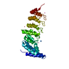 4hbdS S: Starting model for refinement |
|---|---|
| Similar structure data |


-Links
 PDBj
PDBj

- Assembly
| Deposited unit | 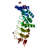
| |||||||||
|---|---|---|---|---|---|---|---|---|---|---|
| 1 |
| |||||||||
| Unit cell |
| |||||||||
| Components on special symmetry positions |
|
-Components
| #1: Protein | Mass: 27942.971 Da / Num. of mol.: 1 / Mutation: Yes; A670V Source method: isolated from a genetically manipulated source Details: residues 583-832 / Source: (gene. exp.) Homo sapiens (human) / Gene: KANK2, ANKRD25, KIAA1518, MXRA3, SIP / Plasmid: PET-151-TOPO / Production host:  Escherichia coli BL21(DE3) (bacteria) / References: UniProt: Q63ZY3 Escherichia coli BL21(DE3) (bacteria) / References: UniProt: Q63ZY3 | ||||||
|---|---|---|---|---|---|---|---|
| #2: Chemical | Glycerol  Mass: 92.094 Da / Num. of mol.: 3 / Source method: obtained synthetically / Formula: C3H8O3 Mass: 92.094 Da / Num. of mol.: 3 / Source method: obtained synthetically / Formula: C3H8O3#3: Chemical | ChemComp-ACT / Acetate  Mass: 59.044 Da / Num. of mol.: 5 / Source method: obtained synthetically / Formula: C2H3O2 Mass: 59.044 Da / Num. of mol.: 5 / Source method: obtained synthetically / Formula: C2H3O2#4: Water | ChemComp-HOH / | Water Mass: 18.015 Da / Num. of mol.: 186 / Source method: isolated from a natural source / Formula: H2O Mass: 18.015 Da / Num. of mol.: 186 / Source method: isolated from a natural source / Formula: H2OHas ligand of interest | N | |
-Experimental details
-Experiment
| Experiment | Method: X-RAY DIFFRACTION / Number of used crystals: 1 |
|---|
- Sample preparation
| Crystal | Density Matthews: 1.97 Å3/Da / Density % sol: 37.53 % |
|---|---|
| Crystal grow | Temperature: 294 K / Method: vapor diffusion, hanging drop / pH: 5.7 Details: 0.2 M ammonium acetate, 0.1 M Bis-tris pH 5.7, 31% w/v PEG 3350 |
-Data collection
| Diffraction | Mean temperature: 100 K / Serial crystal experiment: N |
|---|---|
| Diffraction source | Source: SYNCHROTRON / Site: Diamond / Beamline: I04-1 / Wavelength: 0.91587 Å |
| Detector | Type: DECTRIS PILATUS 6M-F / Detector: PIXEL / Date: Jun 28, 2019 |
| Radiation | Protocol: SINGLE WAVELENGTH / Monochromatic (M) / Laue (L): M / Scattering type: x-ray |
| Radiation wavelength | Wavelength: 0.91587 Å / Relative weight: 1 |
| Reflection | Resolution: 1.5→47.15 Å / Num. obs: 35000 / % possible obs: 99.2 % / Redundancy: 3.4 % / CC1/2: 0.999 / Rmerge(I) obs: 0.056 / Rpim(I) all: 0.036 / Rrim(I) all: 0.067 / Net I/σ(I): 10.3 |
| Reflection shell | Resolution: 1.5→1.521 Å / Redundancy: 3.2 % / Rmerge(I) obs: 2.239 / Mean I/σ(I) obs: 0.5 / Num. unique obs: 1775 / CC1/2: 0.329 / Rpim(I) all: 1.45 / Rrim(I) all: 2.674 / % possible all: 99.9 |
- Processing
| Software |
| ||||||||||||||||||||||||||||||||||||||||||||||||||||||||||||
|---|---|---|---|---|---|---|---|---|---|---|---|---|---|---|---|---|---|---|---|---|---|---|---|---|---|---|---|---|---|---|---|---|---|---|---|---|---|---|---|---|---|---|---|---|---|---|---|---|---|---|---|---|---|---|---|---|---|---|---|---|---|
| Refinement | Method to determine structure: MOLECULAR REPLACEMENT Starting model: 4hbd Resolution: 1.5→47.15 Å / Cor.coef. Fo:Fc: 0.977 / Cor.coef. Fo:Fc free: 0.964 / SU B: 3.194 / SU ML: 0.1 / Cross valid method: THROUGHOUT / σ(F): 0 / ESU R: 0.08 / ESU R Free: 0.086 Details: HYDROGENS HAVE BEEN ADDED IN THE RIDING POSITIONS U VALUES : REFINED INDIVIDUALLY
| ||||||||||||||||||||||||||||||||||||||||||||||||||||||||||||
| Solvent computation | Ion probe radii: 0.8 Å / Shrinkage radii: 0.8 Å / VDW probe radii: 1.2 Å | ||||||||||||||||||||||||||||||||||||||||||||||||||||||||||||
| Displacement parameters | Biso max: 86.95 Å2 / Biso mean: 27.083 Å2 / Biso min: 17.23 Å2
| ||||||||||||||||||||||||||||||||||||||||||||||||||||||||||||
| Refinement step | Cycle: final / Resolution: 1.5→47.15 Å
| ||||||||||||||||||||||||||||||||||||||||||||||||||||||||||||
| Refine LS restraints |
| ||||||||||||||||||||||||||||||||||||||||||||||||||||||||||||
| LS refinement shell | Resolution: 1.5→1.534 Å / Rfactor Rfree error: 0
|
