 Movie
Movie Controller

[English] 日本語
 Yorodumi
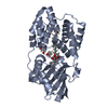
- PDB-6t6k: Y201W mutant of the orange carotenoid protein from Synechocystis ... -
+ Open data

- Basic information
| Entry | Database: PDB / ID: 6t6k | ||||||
|---|---|---|---|---|---|---|---|
| Title | Y201W mutant of the orange carotenoid protein from Synechocystis at pH 6.5 | ||||||
 Components Components | Orange carotenoid-binding protein | ||||||
 Keywords Keywords |  PLANT PROTEIN / orange carotenoid protein / photoactive protein / mutant PLANT PROTEIN / orange carotenoid protein / photoactive protein / mutant | ||||||
| Function / homology |  Function and homology informationlight absorption / phycobilisome / chloride ion binding / plasma membrane-derived thylakoid membrane / photoreceptor activity Function and homology informationlight absorption / phycobilisome / chloride ion binding / plasma membrane-derived thylakoid membrane / photoreceptor activitySimilarity search - Function | ||||||
| Biological species |  | ||||||
| Method | X-RAY DIFFRACTION / SYNCHROTRON / MOLECULAR REPLACEMENT / Resolution: 1.37 Å | ||||||
 Authors Authors | Sluchanko, N.N. / Gushchin, I. / Botnarevskiy, V.S. / Slonimskiy, Y.B. / Remeeva, A. / Kovalev, K. / Stepanov, A.V. / Gordeliy, V. / Maksimov, E.G. | ||||||
| Funding support |  Russian Federation, 1items Russian Federation, 1items
| ||||||
 Citation Citation | Journal: Commun Biol / Year: 2021 Title: Role of hydrogen bond alternation and charge transfer states in photoactivation of the Orange Carotenoid Protein. Authors: Yaroshevich, I.A. / Maksimov, E.G. / Sluchanko, N.N. / Zlenko, D.V. / Stepanov, A.V. / Slutskaya, E.A. / Slonimskiy, Y.B. / Botnarevskii, V.S. / Remeeva, A. / Gushchin, I. / Kovalev, K. / ...Authors: Yaroshevich, I.A. / Maksimov, E.G. / Sluchanko, N.N. / Zlenko, D.V. / Stepanov, A.V. / Slutskaya, E.A. / Slonimskiy, Y.B. / Botnarevskii, V.S. / Remeeva, A. / Gushchin, I. / Kovalev, K. / Gordeliy, V.I. / Shelaev, I.V. / Gostev, F.E. / Khakhulin, D. / Poddubnyy, V.V. / Gostev, T.S. / Cherepanov, D.A. / Polivka, T. / Kloz, M. / Friedrich, T. / Paschenko, V.Z. / Nadtochenko, V.A. / Rubin, A.B. / Kirpichnikov, M.P. | ||||||
| History |
|
- Structure visualization
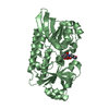
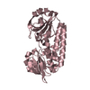
| Structure viewer | Molecule: MolmilJmol/JSmol |
|---|
- Downloads & links
-Download
| PDBx/mmCIF format | 6t6k.cif.gz | 159.2 KB | Display | PDBx/mmCIF format |
|---|---|---|---|---|
| PDB format | pdb6t6k.ent.gz | 122.4 KB | Display | PDB format |
| PDBx/mmJSON format | 6t6k.json.gz | Tree view | PDBx/mmJSON format | |
| Others |  Other downloads Other downloads |
-Validation report
| Arichive directory | https://data.pdbj.org/pub/pdb/validation_reports/t6/6t6kftp://data.pdbj.org/pub/pdb/validation_reports/t6/6t6k | HTTPS FTP |
|---|
-Related structure data
| Related structure data |  6t6mC  6t6oC  4xb5S S: Starting model for refinement C: citing same article ( |
|---|---|
| Similar structure data |


-Links
 PDBj
PDBj


- Assembly
| Deposited unit | 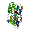
| ||||||||
|---|---|---|---|---|---|---|---|---|---|
| 1 |
| ||||||||
| Unit cell |
|
-Components
-Protein , 1 types, 1 molecules A
| #1: Protein | Mass: 36454.523 Da / Num. of mol.: 1 Source method: isolated from a genetically manipulated source Source: (gene. exp.) Strain: PCC 6803 / Kazusa / Gene: slr1963 / Production host: Escherichia coli (E. coli) / References: UniProt: P74102 |
|---|
-Non-polymers , 7 types, 312 molecules 


| #2: Chemical | ChemComp-ECH / Echinenone Mass: 550.856 Da / Num. of mol.: 1 / Source method: obtained synthetically / Formula: C40H54O / Feature type: SUBJECT OF INVESTIGATION Mass: 550.856 Da / Num. of mol.: 1 / Source method: obtained synthetically / Formula: C40H54O / Feature type: SUBJECT OF INVESTIGATION | ||||||||||
|---|---|---|---|---|---|---|---|---|---|---|---|
| #3: Chemical | ChemComp-GLY / Glycine Type: peptide linking / Mass: 75.067 Da / Num. of mol.: 5 / Source method: obtained synthetically / Formula: C2H5NO2 Type: peptide linking / Mass: 75.067 Da / Num. of mol.: 5 / Source method: obtained synthetically / Formula: C2H5NO2#4: Chemical | ChemComp-CL / | Chloride Mass: 35.453 Da / Num. of mol.: 1 / Source method: obtained synthetically / Formula: Cl Mass: 35.453 Da / Num. of mol.: 1 / Source method: obtained synthetically / Formula: Cl#5: Chemical | ChemComp-IMD / | Imidazole Mass: 69.085 Da / Num. of mol.: 1 / Source method: obtained synthetically / Formula: C3H5N2 Mass: 69.085 Da / Num. of mol.: 1 / Source method: obtained synthetically / Formula: C3H5N2#6: Chemical | ChemComp-HIS / | Histidine Type: L-peptide linking / Mass: 156.162 Da / Num. of mol.: 1 / Source method: obtained synthetically / Formula: C6H10N3O2 Type: L-peptide linking / Mass: 156.162 Da / Num. of mol.: 1 / Source method: obtained synthetically / Formula: C6H10N3O2#7: Chemical | ChemComp-EDO / | Ethylene glycol Mass: 62.068 Da / Num. of mol.: 1 / Source method: obtained synthetically / Formula: C2H6O2 Mass: 62.068 Da / Num. of mol.: 1 / Source method: obtained synthetically / Formula: C2H6O2#8: Water | ChemComp-HOH / | WaterMass: 18.015 Da / Num. of mol.: 302 / Source method: isolated from a natural source / Formula: H2O |
-Details
| Has ligand of interest | Y |
|---|
-Experimental details
-Experiment
| Experiment | Method: X-RAY DIFFRACTION / Number of used crystals: 1 |
|---|
- Sample preparation
| Crystal | Density Matthews: 2.39 Å3/Da / Density % sol: 48.44 % |
|---|---|
| Crystal grow | Temperature: 293 K / Method: vapor diffusion / pH: 6.5 Details: 0.1M DL-Glutamic acid monohydrate; 0.1M DL-Alanine; 0.1M Glycine; 0.1M DL-Lysine monohydrochloride; 0.1M DL-Serine; 0.1M Imidazole; MES monohydrate (acid), pH 6.5; 20% v/v Ethylene glycol; 10 % w/v PEG 8000 |
-Data collection
| Diffraction | Mean temperature: 100 K / Serial crystal experiment: N | ||||||||||||||||||||||||
|---|---|---|---|---|---|---|---|---|---|---|---|---|---|---|---|---|---|---|---|---|---|---|---|---|---|
| Diffraction source | Source: SYNCHROTRON / Site: PETRA III, EMBL c/o DESY  / Beamline: P14 (MX2) / Wavelength: 0.97625 Å / Beamline: P14 (MX2) / Wavelength: 0.97625 Å | ||||||||||||||||||||||||
| Detector | Type: DECTRIS EIGER X 16M / Detector: PIXEL / Date: Oct 6, 2019 | ||||||||||||||||||||||||
| Radiation | Protocol: SINGLE WAVELENGTH / Monochromatic (M) / Laue (L): M / Scattering type: x-ray | ||||||||||||||||||||||||
| Radiation wavelength | Wavelength: 0.97625 Å / Relative weight: 1 | ||||||||||||||||||||||||
| Reflection | Resolution: 1.37→44.02 Å / Num. obs: 73479 / % possible obs: 100 % / Redundancy: 20.2 % / CC1/2: 0.999 / Rmerge(I) obs: 0.047 / Net I/σ(I): 29.1 / Num. measured all: 1480976 / Scaling rejects: 14 | ||||||||||||||||||||||||
| Reflection shell | Diffraction-ID: 1
|
- Processing
| Software |
| |||||||||||||||||||||||||||||||||||||||||||||||||||||||||||||||||||||||||||
|---|---|---|---|---|---|---|---|---|---|---|---|---|---|---|---|---|---|---|---|---|---|---|---|---|---|---|---|---|---|---|---|---|---|---|---|---|---|---|---|---|---|---|---|---|---|---|---|---|---|---|---|---|---|---|---|---|---|---|---|---|---|---|---|---|---|---|---|---|---|---|---|---|---|---|---|---|
| Refinement | Method to determine structure: MOLECULAR REPLACEMENT Starting model: 4XB5 Resolution: 1.37→37.54 Å / Cor.coef. Fo:Fc: 0.981 / Cor.coef. Fo:Fc free: 0.974 / SU B: 2.263 / SU ML: 0.039 / Cross valid method: THROUGHOUT / σ(F): 0 / ESU R: 0.048 / ESU R Free: 0.047 Details: HYDROGENS HAVE BEEN ADDED IN THE RIDING POSITIONS U VALUES : WITH TLS ADDED
| |||||||||||||||||||||||||||||||||||||||||||||||||||||||||||||||||||||||||||
| Solvent computation | Ion probe radii: 0.8 Å / Shrinkage radii: 0.8 Å / VDW probe radii: 1.2 Å | |||||||||||||||||||||||||||||||||||||||||||||||||||||||||||||||||||||||||||
| Displacement parameters | Biso max: 205.74 Å2 / Biso mean: 29.765 Å2 / Biso min: 17.15 Å2
| |||||||||||||||||||||||||||||||||||||||||||||||||||||||||||||||||||||||||||
| Refinement step | Cycle: final / Resolution: 1.37→37.54 Å
| |||||||||||||||||||||||||||||||||||||||||||||||||||||||||||||||||||||||||||
| Refine LS restraints |
| |||||||||||||||||||||||||||||||||||||||||||||||||||||||||||||||||||||||||||
| LS refinement shell | Resolution: 1.37→1.406 Å / Rfactor Rfree error: 0 / Total num. of bins used: 20
| |||||||||||||||||||||||||||||||||||||||||||||||||||||||||||||||||||||||||||
| Refinement TLS params. | Method: refined / Refine-ID: X-RAY DIFFRACTION
| |||||||||||||||||||||||||||||||||||||||||||||||||||||||||||||||||||||||||||
| Refinement TLS group |
|
