 Movie
Movie Controller

[English] 日本語
 Yorodumi
- PDB-6swx: Leishmania major methionyl-tRNA synthetase in complex with an all... -
+ Open data

- Basic information
| Entry | Database: PDB / ID: 6swx | ||||||
|---|---|---|---|---|---|---|---|
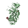
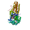
| Title | Leishmania major methionyl-tRNA synthetase in complex with an allosteric inhibitor | ||||||
 Components Components | Putative methionyl-tRNA synthetase Methionine—tRNA ligase Methionine—tRNA ligase | ||||||
 Keywords Keywords | TRANSLATION / LEISHMANIA / PARASITE / AMINOACYL-TRNA SYNTHETASE / TRNA LIGASE / AARS / METRS / METHIONINE / ATP-BINDING / NUCLEOTIDE-BINDING / LIGASE / DRUG DISCOVERY / DUNDEE DRUG DISCOVERY UNIT | ||||||
| Function / homology |  Function and homology informationmethionine-tRNA ligase / methionine-tRNA ligase activity / methionyl-tRNA aminoacylation / ciliary plasm / magnesium ion binding / ATP binding / cytoplasm Function and homology informationmethionine-tRNA ligase / methionine-tRNA ligase activity / methionyl-tRNA aminoacylation / ciliary plasm / magnesium ion binding / ATP binding / cytoplasmSimilarity search - Function | ||||||
| Biological species |  Leishmania major (eukaryote) Leishmania major (eukaryote) | ||||||
| Method | X-RAY DIFFRACTION / SYNCHROTRON / MOLECULAR REPLACEMENT / Resolution: 1.95 Å | ||||||
 Authors Authors | Robinson, D.A. / Torrie, L.S. / Shepherd, S.M. / De Rycker, M. / Thomas, M.G. / Wyatt, P.G. | ||||||
| Funding support |  United Kingdom, 1items United Kingdom, 1items
| ||||||
 Citation Citation | Journal: Acs Infect Dis. / Year: 2020 Title: Discovery of an Allosteric Binding Site in Kinetoplastid Methionyl-tRNA Synthetase. Authors: Torrie, L.S. / Robinson, D.A. / Thomas, M.G. / Hobrath, J.V. / Shepherd, S.M. / Post, J.M. / Ko, E.J. / Ferreira, R.A. / Mackenzie, C.J. / Wrobel, K. / Edwards, D.P. / Gilbert, I.H. / Gray, ...Authors: Torrie, L.S. / Robinson, D.A. / Thomas, M.G. / Hobrath, J.V. / Shepherd, S.M. / Post, J.M. / Ko, E.J. / Ferreira, R.A. / Mackenzie, C.J. / Wrobel, K. / Edwards, D.P. / Gilbert, I.H. / Gray, D.W. / Fairlamb, A.H. / De Rycker, M. | ||||||
| History |
|
- Structure visualization
| Structure viewer | Molecule: MolmilJmol/JSmol |
|---|
- Downloads & links
-Download
| PDBx/mmCIF format | 6swx.cif.gz | 130.9 KB | Display | PDBx/mmCIF format |
|---|---|---|---|---|
| PDB format | pdb6swx.ent.gz | 97.9 KB | Display | PDB format |
| PDBx/mmJSON format | 6swx.json.gz | Tree view | PDBx/mmJSON format | |
| Others |  Other downloads Other downloads |
-Validation report
| Arichive directory | https://data.pdbj.org/pub/pdb/validation_reports/sw/6swxftp://data.pdbj.org/pub/pdb/validation_reports/sw/6swx | HTTPS FTP |
|---|
-Related structure data
| Related structure data |  3kflS S: Starting model for refinement |
|---|---|
| Similar structure data |


-Links
 PDBj
PDBj
- Assembly
| Deposited unit | 
| ||||||||
|---|---|---|---|---|---|---|---|---|---|
| 1 |
| ||||||||
| Unit cell |
|
-Components
| #1: Protein | Methionine—tRNA ligase Mass: 63984.793 Da / Num. of mol.: 1 Source method: isolated from a genetically manipulated source Source: (gene. exp.) Leishmania major (eukaryote) / Gene: LMJF_21_0810 / Production host:  Escherichia coli BL21 (bacteria) / References: UniProt: Q4QCD2, methionine-tRNA ligase Escherichia coli BL21 (bacteria) / References: UniProt: Q4QCD2, methionine-tRNA ligase |
|---|---|
| #2: Chemical | ChemComp-LWN /   Mass: 348.307 Da / Num. of mol.: 1 / Source method: obtained synthetically / Formula: C15H14F2N6O2 / Feature type: SUBJECT OF INVESTIGATION Mass: 348.307 Da / Num. of mol.: 1 / Source method: obtained synthetically / Formula: C15H14F2N6O2 / Feature type: SUBJECT OF INVESTIGATION |
| #3: Chemical | ChemComp-MET / Methionine  Type: L-peptide linking / Mass: 149.211 Da / Num. of mol.: 1 / Source method: obtained synthetically / Formula: C5H11NO2S Type: L-peptide linking / Mass: 149.211 Da / Num. of mol.: 1 / Source method: obtained synthetically / Formula: C5H11NO2S |
| #4: Water | ChemComp-HOH / Water Mass: 18.015 Da / Num. of mol.: 326 / Source method: isolated from a natural source / Formula: H2O Mass: 18.015 Da / Num. of mol.: 326 / Source method: isolated from a natural source / Formula: H2O |
| Has ligand of interest | Y |
-Experimental details
-Experiment
| Experiment | Method: X-RAY DIFFRACTION / Number of used crystals: 1 |
|---|
- Sample preparation
| Crystal | Density Matthews: 2.99 Å3/Da / Density % sol: 58.84 % / Description: Large Cuboid |
|---|---|
| Crystal grow | Temperature: 291 K / Method: vapor diffusion, sitting drop / Details: 23-28% PEG 3350, 0.2 M K Formate pH 7.0-7.5 / PH range: 7.0 - 7.5 |
-Data collection
| Diffraction | Mean temperature: 100 K / Serial crystal experiment: N |
|---|---|
| Diffraction source | Source: SYNCHROTRON / Site: Diamond / Beamline: I03 / Wavelength: 0.976 Å |
| Detector | Type: DECTRIS PILATUS 6M / Detector: PIXEL / Date: May 14, 2017 |
| Radiation | Protocol: SINGLE WAVELENGTH / Monochromatic (M) / Laue (L): M / Scattering type: x-ray |
| Radiation wavelength | Wavelength: 0.976 Å / Relative weight: 1 |
| Reflection | Resolution: 1.95→28.55 Å / Num. obs: 53240 / % possible obs: 99.8 % / Redundancy: 6.5 % / Biso Wilson estimate: 35 Å2 / CC1/2: 1 / Rmerge(I) obs: 0.037 / Net I/σ(I): 24 |
| Reflection shell | Resolution: 1.95→2 Å / Redundancy: 6.6 % / Rmerge(I) obs: 0.711 / Mean I/σ(I) obs: 2.4 / Num. unique obs: 3650 / CC1/2: 0.87 / % possible all: 98 |
- Processing
| Software |
| |||||||||||||||||||||||||||||||||||||||||||||||||||||||||||||||||||||||||||
|---|---|---|---|---|---|---|---|---|---|---|---|---|---|---|---|---|---|---|---|---|---|---|---|---|---|---|---|---|---|---|---|---|---|---|---|---|---|---|---|---|---|---|---|---|---|---|---|---|---|---|---|---|---|---|---|---|---|---|---|---|---|---|---|---|---|---|---|---|---|---|---|---|---|---|---|---|
| Refinement | Method to determine structure: MOLECULAR REPLACEMENT Starting model: 3KFL Resolution: 1.95→28.55 Å / Cor.coef. Fo:Fc: 0.968 / Cor.coef. Fo:Fc free: 0.952 / Cross valid method: THROUGHOUT / σ(F): 0 / ESU R: 0.14 / ESU R Free: 0.128 Details: HYDROGENS HAVE BEEN ADDED IN THE RIDING POSITIONS U VALUES : REFINED INDIVIDUALLY
| |||||||||||||||||||||||||||||||||||||||||||||||||||||||||||||||||||||||||||
| Solvent computation | Ion probe radii: 0.8 Å / Shrinkage radii: 0.8 Å / VDW probe radii: 1.2 Å | |||||||||||||||||||||||||||||||||||||||||||||||||||||||||||||||||||||||||||
| Displacement parameters | Biso max: 167.72 Å2 / Biso mean: 45.156 Å2 / Biso min: 26.22 Å2
| |||||||||||||||||||||||||||||||||||||||||||||||||||||||||||||||||||||||||||
| Refinement step | Cycle: final / Resolution: 1.95→28.55 Å
| |||||||||||||||||||||||||||||||||||||||||||||||||||||||||||||||||||||||||||
| Refine LS restraints |
| |||||||||||||||||||||||||||||||||||||||||||||||||||||||||||||||||||||||||||
| LS refinement shell | Resolution: 1.954→2.004 Å / Total num. of bins used: 20
|
