 Movie
Movie Controller

[English] 日本語
 Yorodumi
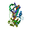
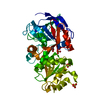
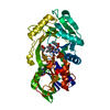
- PDB-6sw2: Crystal Structure of P. aeruginosa PqsL in complex with 2-aminobe... -
+ Open data

- Basic information
| Entry | Database: PDB / ID: 6sw2 | ||||||
|---|---|---|---|---|---|---|---|
| Title | Crystal Structure of P. aeruginosa PqsL in complex with 2-aminobenzoylacetate | ||||||
 Components Components | Probable FAD-dependent monooxygenase | ||||||
 Keywords Keywords |  ANTIBIOTIC / oxidoreductase / photocatalysis / FAD / NAD(P) / biosynthetic pathway ANTIBIOTIC / oxidoreductase / photocatalysis / FAD / NAD(P) / biosynthetic pathway | ||||||
| Function / homology |  Function and homology information Function and homology information3-(3-hydroxyphenyl)propionate hydroxylase activity / 3-(3-hydroxy)phenylpropionate catabolic process / FAD binding Similarity search - Function | ||||||
| Biological species |   Pseudomonas aeruginosa (bacteria) Pseudomonas aeruginosa (bacteria) | ||||||
| Method | X-RAY DIFFRACTION / SYNCHROTRON / MOLECULAR REPLACEMENT / molecular replacement / Resolution: 1.7 Å | ||||||
 Authors Authors | Mattevi, A. / Rovida, S. | ||||||
 Citation Citation | Journal: Nat Commun / Year: 2020 Title: Photoinduced monooxygenation involving NAD(P)H-FAD sequential single-electron transfer. Authors: Ernst, S. / Rovida, S. / Mattevi, A. / Fetzner, S. / Drees, S.L. | ||||||
| History |
|
- Structure visualization
| Structure viewer | Molecule: MolmilJmol/JSmol |
|---|
- Downloads & links
-Download
| PDBx/mmCIF format | 6sw2.cif.gz | 98.4 KB | Display | PDBx/mmCIF format |
|---|---|---|---|---|
| PDB format | pdb6sw2.ent.gz | 71.2 KB | Display | PDB format |
| PDBx/mmJSON format | 6sw2.json.gz | Tree view | PDBx/mmJSON format | |
| Others |  Other downloads Other downloads |
-Validation report
| Arichive directory | https://data.pdbj.org/pub/pdb/validation_reports/sw/6sw2ftp://data.pdbj.org/pub/pdb/validation_reports/sw/6sw2 | HTTPS FTP |
|---|
-Related structure data
| Related structure data |  6sw1C 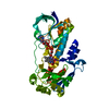 2x3nS S: Starting model for refinement C: citing same article ( |
|---|---|
| Similar structure data |


-Links
 PDBj
PDBj- Assembly
| Deposited unit | 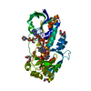
| ||||||||
|---|---|---|---|---|---|---|---|---|---|
| 1 |
| ||||||||
| Unit cell |
|
-Components
-Protein , 1 types, 1 molecules A
| #1: Protein | Mass: 44047.973 Da / Num. of mol.: 1 Source method: isolated from a genetically manipulated source Source: (gene. exp.) Pseudomonas aeruginosa (strain ATCC 15692 / DSM 22644 / CIP 104116 / JCM 14847 / LMG 12228 / 1C / PRS 101 / PAO1) (bacteria)Gene: pqsL, PA4190 / Production host: Escherichia coli (E. coli) / Strain (production host): LMG194 / References: UniProt: Q9HWJ1 |
|---|
-Non-polymers , 7 types, 185 molecules 


| #2: Chemical | ChemComp-FAD / Flavin adenine dinucleotide Mass: 785.550 Da / Num. of mol.: 1 / Source method: obtained synthetically / Formula: C27H33N9O15P2 / Feature type: SUBJECT OF INVESTIGATION / Comment: FAD*YM Mass: 785.550 Da / Num. of mol.: 1 / Source method: obtained synthetically / Formula: C27H33N9O15P2 / Feature type: SUBJECT OF INVESTIGATION / Comment: FAD*YM | ||||||||
|---|---|---|---|---|---|---|---|---|---|
| #3: Chemical | ChemComp-61M /  Mass: 179.173 Da / Num. of mol.: 1 / Source method: obtained synthetically / Formula: C9H9NO3 / Feature type: SUBJECT OF INVESTIGATION Mass: 179.173 Da / Num. of mol.: 1 / Source method: obtained synthetically / Formula: C9H9NO3 / Feature type: SUBJECT OF INVESTIGATION | ||||||||
| #4: Chemical | ChemComp-PGE / Polyethylene glycol Mass: 150.173 Da / Num. of mol.: 4 / Source method: obtained synthetically / Formula: C6H14O4 / Feature type: SUBJECT OF INVESTIGATION Mass: 150.173 Da / Num. of mol.: 4 / Source method: obtained synthetically / Formula: C6H14O4 / Feature type: SUBJECT OF INVESTIGATION#5: Chemical | ChemComp-PO4 / | Phosphate Mass: 94.971 Da / Num. of mol.: 1 / Source method: obtained synthetically / Formula: PO4 Mass: 94.971 Da / Num. of mol.: 1 / Source method: obtained synthetically / Formula: PO4#6: Chemical | ChemComp-MES / | MES (buffer) Mass: 195.237 Da / Num. of mol.: 1 / Source method: obtained synthetically / Formula: C6H13NO4S / Comment: pH buffer*YM Mass: 195.237 Da / Num. of mol.: 1 / Source method: obtained synthetically / Formula: C6H13NO4S / Comment: pH buffer*YM#7: Chemical | Diethylene glycol Mass: 106.120 Da / Num. of mol.: 2 / Source method: obtained synthetically / Formula: C4H10O3 Mass: 106.120 Da / Num. of mol.: 2 / Source method: obtained synthetically / Formula: C4H10O3#8: Water | ChemComp-HOH / | WaterMass: 18.015 Da / Num. of mol.: 175 / Source method: isolated from a natural source / Formula: H2O |
-Details
| Has ligand of interest | Y |
|---|
-Experimental details
-Experiment
| Experiment | Method: X-RAY DIFFRACTION / Number of used crystals: 1 |
|---|
- Sample preparation
| Crystal | Density Matthews: 2.19 Å3/Da / Density % sol: 43.86 % |
|---|---|
| Crystal grow | Temperature: 298 K / Method: vapor diffusion, sitting drop / pH: 6.5 / Details: 20,000 (5-10 % w/v), MES buffer pH 6.5. |
-Data collection
| Diffraction | Mean temperature: 100 K / Serial crystal experiment: N | ||||||||||||||||||||||||||||||
|---|---|---|---|---|---|---|---|---|---|---|---|---|---|---|---|---|---|---|---|---|---|---|---|---|---|---|---|---|---|---|---|
| Diffraction source | Source: SYNCHROTRON / Site: ESRF  / Beamline: ID23-1 / Wavelength: 0.966 Å / Beamline: ID23-1 / Wavelength: 0.966 Å | ||||||||||||||||||||||||||||||
| Detector | Type: DECTRIS PILATUS3 S 6M / Detector: PIXEL / Date: Apr 3, 2018 | ||||||||||||||||||||||||||||||
| Radiation | Protocol: SINGLE WAVELENGTH / Monochromatic (M) / Laue (L): M / Scattering type: x-ray | ||||||||||||||||||||||||||||||
| Radiation wavelength | Wavelength: 0.966 Å / Relative weight: 1 | ||||||||||||||||||||||||||||||
| Reflection | Resolution: 1.7→45.31 Å / Num. obs: 43115 / % possible obs: 99.3 % / Redundancy: 4.8 % / CC1/2: 0.998 / Rmerge(I) obs: 0.075 / Rpim(I) all: 0.037 / Rrim(I) all: 0.085 / Net I/σ(I): 10.1 | ||||||||||||||||||||||||||||||
| Reflection shell | Diffraction-ID: 1
|
-Phasing
| Phasing | Method: molecular replacement |
|---|
- Processing
| Software |
| ||||||||||||||||||||||||||||||||||||||||||||||||||||||||||||
|---|---|---|---|---|---|---|---|---|---|---|---|---|---|---|---|---|---|---|---|---|---|---|---|---|---|---|---|---|---|---|---|---|---|---|---|---|---|---|---|---|---|---|---|---|---|---|---|---|---|---|---|---|---|---|---|---|---|---|---|---|---|
| Refinement | Method to determine structure: MOLECULAR REPLACEMENT Starting model: 2X3N Resolution: 1.7→45.31 Å / Cor.coef. Fo:Fc: 0.97 / Cor.coef. Fo:Fc free: 0.953 / SU B: 2.769 / SU ML: 0.085 / Cross valid method: THROUGHOUT / σ(F): 0 / ESU R: 0.101 / ESU R Free: 0.105 Details: HYDROGENS HAVE BEEN ADDED IN THE RIDING POSITIONS U VALUES - REFINED INDIVIDUALLY
| ||||||||||||||||||||||||||||||||||||||||||||||||||||||||||||
| Solvent computation | Ion probe radii: 0.8 Å / Shrinkage radii: 0.8 Å / VDW probe radii: 1.2 Å | ||||||||||||||||||||||||||||||||||||||||||||||||||||||||||||
| Displacement parameters | Biso max: 92.86 Å2 / Biso mean: 29.736 Å2 / Biso min: 16.27 Å2
| ||||||||||||||||||||||||||||||||||||||||||||||||||||||||||||
| Refinement step | Cycle: final / Resolution: 1.7→45.31 Å
| ||||||||||||||||||||||||||||||||||||||||||||||||||||||||||||
| Refine LS restraints |
| ||||||||||||||||||||||||||||||||||||||||||||||||||||||||||||
| LS refinement shell | Resolution: 1.7→1.744 Å / Rfactor Rfree error: 0 / Total num. of bins used: 20
|
