 Movie
Movie Controller


+ Open data

- Basic information
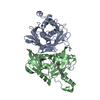
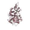
| Entry | Database: PDB / ID: 6sqj | ||||||
|---|---|---|---|---|---|---|---|
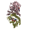
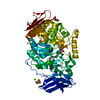
| Title | Crystal structure of glycoprotein D of Equine Herpesvirus Type 1 | ||||||
 Components Components | Glycoprotein D | ||||||
 Keywords Keywords |  VIRAL PROTEIN / glycoprotein D / equine herpesvirus type 1 / viral entry VIRAL PROTEIN / glycoprotein D / equine herpesvirus type 1 / viral entry | ||||||
| Function / homology | Herpesvirus glycoprotein D/GG/GX domain / Herpesvirus glycoprotein D/GG/GX domain / membrane => GO:0016020 / Immunoglobulin-like domain superfamily / viral envelope / Glycoprotein D / Envelope glycoprotein D Function and homology information Function and homology information | ||||||
| Biological species |  Equid alphaherpesvirus 1 (Equine herpesvirus 1) Equid alphaherpesvirus 1 (Equine herpesvirus 1) | ||||||
| Method | X-RAY DIFFRACTION / SYNCHROTRON / MOLECULAR REPLACEMENT / Resolution: 2.245 Å | ||||||
 Authors Authors | Kremling, V. / Loll, B. / Azab, W. / Osterrieder, N. / Dahmani, I. / Chiantia, P. / Wahl, M. | ||||||
| Funding support |  Germany, 1items Germany, 1items
| ||||||
 Citation Citation | Journal: Front Microbiol / Year: 2023 Title: Crystal structures of glycoprotein D of equine alphaherpesviruses reveal potential binding sites to the entry receptor MHC-I. Authors: Kremling, V. / Loll, B. / Pach, S. / Dahmani, I. / Weise, C. / Wolber, G. / Chiantia, S. / Wahl, M.C. / Osterrieder, N. / Azab, W. | ||||||
| History |
|
- Structure visualization
| Structure viewer | Molecule: MolmilJmol/JSmol |
|---|
- Downloads & links
-Download
| PDBx/mmCIF format | 6sqj.cif.gz | 117.2 KB | Display | PDBx/mmCIF format |
|---|---|---|---|---|
| PDB format | pdb6sqj.ent.gz | 87.6 KB | Display | PDB format |
| PDBx/mmJSON format | 6sqj.json.gz | Tree view | PDBx/mmJSON format | |
| Others |  Other downloads Other downloads |
-Validation report
| Arichive directory | https://data.pdbj.org/pub/pdb/validation_reports/sq/6sqjftp://data.pdbj.org/pub/pdb/validation_reports/sq/6sqj | HTTPS FTP |
|---|
-Related structure data
| Related structure data | 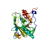 6tm8C 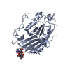 2c36S S: Starting model for refinement C: citing same article ( |
|---|---|
| Similar structure data |


-Links
 PDBj
PDBj- Assembly
| Deposited unit | 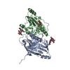
| ||||||||
|---|---|---|---|---|---|---|---|---|---|
| 1 |
| ||||||||
| Unit cell |
|
-Components
| #1: Protein | Mass: 37990.988 Da / Num. of mol.: 2 Source method: isolated from a genetically manipulated source Source: (gene. exp.) Equid alphaherpesvirus 1 (Equine herpesvirus 1)Plasmid: pMultiBacY / Cell line (production host): H5 / Production host:  Trichoplusia ni (cabbage looper) / References: UniProt: G9HV37, UniProt: Q04245*PLUS Trichoplusia ni (cabbage looper) / References: UniProt: G9HV37, UniProt: Q04245*PLUS#2: Polysaccharide | / Mass: 424.401 Da / Num. of mol.: 2Source method: isolated from a genetically manipulated source #3: Chemical |   Mass: 24.305 Da / Num. of mol.: 2 / Source method: obtained synthetically / Formula: Mg Mass: 24.305 Da / Num. of mol.: 2 / Source method: obtained synthetically / Formula: Mg#4: Sugar | ChemComp-NAG / | N-Acetylglucosamine  Type: D-saccharide, beta linking / Mass: 221.208 Da / Num. of mol.: 1 Type: D-saccharide, beta linking / Mass: 221.208 Da / Num. of mol.: 1Source method: isolated from a genetically manipulated source Formula: C8H15NO6 #5: Water | ChemComp-HOH / | Water Mass: 18.015 Da / Num. of mol.: 132 / Source method: isolated from a natural source / Formula: H2O Mass: 18.015 Da / Num. of mol.: 132 / Source method: isolated from a natural source / Formula: H2OHas ligand of interest | N | |
|---|
-Experimental details
-Experiment
| Experiment | Method: X-RAY DIFFRACTION / Number of used crystals: 1 |
|---|
- Sample preparation
| Crystal | Density Matthews: 3.18 Å3/Da / Density % sol: 61.36 % |
|---|---|
| Crystal grow | Temperature: 291.15 K / Method: vapor diffusion, sitting drop / pH: 8.5 Details: 0.1 M Tris buffer, 0.2 M magnesium chloride, 30% (w/v) PEG 4000 |
-Data collection
| Diffraction | Mean temperature: 100 K / Serial crystal experiment: N |
|---|---|
| Diffraction source | Source: SYNCHROTRON / Site: PETRA III, DESY / Beamline: P11 / Wavelength: 1.0332 Å |
| Detector | Type: DECTRIS PILATUS 6M / Detector: PIXEL / Date: Oct 26, 2018 |
| Radiation | Protocol: SINGLE WAVELENGTH / Monochromatic (M) / Laue (L): M / Scattering type: x-ray |
| Radiation wavelength | Wavelength: 1.0332 Å / Relative weight: 1 |
| Reflection | Resolution: 2.24→50 Å / Num. obs: 33402 / % possible obs: 99.1 % / Redundancy: 6.5 % / CC1/2: 0.998 / Net I/σ(I): 11.71 |
| Reflection shell | Resolution: 2.24→2.38 Å / Num. unique obs: 5140 / CC1/2: 0.586 |
- Processing
| Software |
| ||||||||||||||||||||||||||||||||||||||||||||||||||||||||||||||||||||||||||||||
|---|---|---|---|---|---|---|---|---|---|---|---|---|---|---|---|---|---|---|---|---|---|---|---|---|---|---|---|---|---|---|---|---|---|---|---|---|---|---|---|---|---|---|---|---|---|---|---|---|---|---|---|---|---|---|---|---|---|---|---|---|---|---|---|---|---|---|---|---|---|---|---|---|---|---|---|---|---|---|---|
| Refinement | Method to determine structure: MOLECULAR REPLACEMENT Starting model: 2C36 Resolution: 2.245→49.803 Å / SU ML: 0.33 / Cross valid method: THROUGHOUT / σ(F): 1.34 / Phase error: 31.73
| ||||||||||||||||||||||||||||||||||||||||||||||||||||||||||||||||||||||||||||||
| Solvent computation | Shrinkage radii: 0.9 Å / VDW probe radii: 1.11 Å | ||||||||||||||||||||||||||||||||||||||||||||||||||||||||||||||||||||||||||||||
| Displacement parameters | Biso max: 142.78 Å2 / Biso mean: 58.7663 Å2 / Biso min: 30.46 Å2 | ||||||||||||||||||||||||||||||||||||||||||||||||||||||||||||||||||||||||||||||
| Refinement step | Cycle: final / Resolution: 2.245→49.803 Å
| ||||||||||||||||||||||||||||||||||||||||||||||||||||||||||||||||||||||||||||||
| LS refinement shell | Refine-ID: X-RAY DIFFRACTION / Rfactor Rfree error: 0
|
