 Movie
Movie Controller

[English] 日本語
 Yorodumi
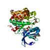
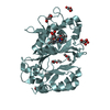
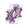
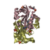
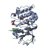
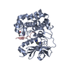
- PDB-6sk1: Diaminobutyrate acetyltransferase EctA from Paenibacillus lautus ... -
+ Open data

- Basic information
| Entry | Database: PDB / ID: 6sk1 | |||||||||
|---|---|---|---|---|---|---|---|---|---|---|
| Title | Diaminobutyrate acetyltransferase EctA from Paenibacillus lautus in complex with coenzyme A | |||||||||
 Components Components | L-2,4-diaminobutyric acid acetyltransferase | |||||||||
 Keywords Keywords |  TRANSFERASE / L-2 / 4-diaminobutyrate acetyltransferase / acetyl coenzyme A / acetylation / stress response / chemical chaperone TRANSFERASE / L-2 / 4-diaminobutyrate acetyltransferase / acetyl coenzyme A / acetylation / stress response / chemical chaperone | |||||||||
| Function / homology |  Function and homology informationdiaminobutyrate acetyltransferase / diaminobutyrate acetyltransferase activity / ectoine biosynthetic process Function and homology informationdiaminobutyrate acetyltransferase / diaminobutyrate acetyltransferase activity / ectoine biosynthetic processSimilarity search - Function | |||||||||
| Biological species |  Geobacillus sp. (bacteria) Geobacillus sp. (bacteria) | |||||||||
| Method | X-RAY DIFFRACTION / SYNCHROTRON / MOLECULAR REPLACEMENT / Resolution: 1.5 Å | |||||||||
 Authors Authors | Richter, A.A. / Kobus, S. / Czech, L. / Hoeppner, A. / Bremer, E. / Smits, S.H.J. | |||||||||
 Citation Citation | Journal: J.Biol.Chem. / Year: 2020 Title: The architecture of the diaminobutyrate acetyltransferase active site provides mechanistic insight into the biosynthesis of the chemical chaperone ectoine. Authors: Richter, A.A. / Kobus, S. / Czech, L. / Hoeppner, A. / Zarzycki, J. / Erb, T.J. / Lauterbach, L. / Dickschat, J.S. / Bremer, E. / Smits, S.H.J. | |||||||||
| History |
|
- Structure visualization
| Structure viewer | Molecule: MolmilJmol/JSmol |
|---|
- Downloads & links
-Download
| PDBx/mmCIF format | 6sk1.cif.gz | 56.9 KB | Display | PDBx/mmCIF format |
|---|---|---|---|---|
| PDB format | pdb6sk1.ent.gz | 38.5 KB | Display | PDB format |
| PDBx/mmJSON format | 6sk1.json.gz | Tree view | PDBx/mmJSON format | |
| Others |  Other downloads Other downloads |
-Validation report
| Arichive directory | https://data.pdbj.org/pub/pdb/validation_reports/sk/6sk1ftp://data.pdbj.org/pub/pdb/validation_reports/sk/6sk1 | HTTPS FTP |
|---|
-Related structure data
| Related structure data |  6sjyC  6sl8C  6slkSC  6sllC S: Starting model for refinement C: citing same article ( |
|---|---|
| Similar structure data |


-Links
 PDBj
PDBj


- Assembly
| Deposited unit | 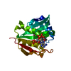
| ||||||||
|---|---|---|---|---|---|---|---|---|---|
| 1 | 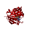
| ||||||||
| Unit cell |
|
-Components
| #1: Protein | Mass: 20827.402 Da / Num. of mol.: 1 Source method: isolated from a genetically manipulated source Source: (gene. exp.) Geobacillus sp. (strain Y412MC10) (bacteria)Strain: Y412MC10 / Gene: ectA, GYMC10_5665 / Production host: Escherichia coli (E. coli)References: UniProt: D3EKC1, diaminobutyrate acetyltransferase |
|---|---|
| #2: Chemical | ChemComp-COA / Coenzyme A  Mass: 767.534 Da / Num. of mol.: 1 / Source method: obtained synthetically / Formula: C21H36N7O16P3S / Feature type: SUBJECT OF INVESTIGATION Mass: 767.534 Da / Num. of mol.: 1 / Source method: obtained synthetically / Formula: C21H36N7O16P3S / Feature type: SUBJECT OF INVESTIGATION |
| #3: Chemical | ChemComp-ACT / Acetate  Mass: 59.044 Da / Num. of mol.: 1 / Source method: isolated from a natural source / Formula: C2H3O2 Mass: 59.044 Da / Num. of mol.: 1 / Source method: isolated from a natural source / Formula: C2H3O2 |
| #4: Water | ChemComp-HOH / Water Mass: 18.015 Da / Num. of mol.: 209 / Source method: isolated from a natural source / Formula: H2O Mass: 18.015 Da / Num. of mol.: 209 / Source method: isolated from a natural source / Formula: H2O |
| Has ligand of interest | Y |
-Experimental details
-Experiment
| Experiment | Method: X-RAY DIFFRACTION / Number of used crystals: 1 |
|---|
- Sample preparation
| Crystal | Density Matthews: 2.58 Å3/Da / Density % sol: 52.41 % |
|---|---|
| Crystal grow | Temperature: 285 K / Method: vapor diffusion, sitting drop Details: 0.1 M magnesium chloride, 0.1 M sodium chloride, 12 % (w/v) PEG 4000, 0.1 M tri-sodium citrate pH 5.5, 5 mM coenzyme A |
-Data collection
| Diffraction | Mean temperature: 100 K / Serial crystal experiment: N |
|---|---|
| Diffraction source | Source: SYNCHROTRON / Site: ESRF  / Beamline: ID29 / Wavelength: 0.9762 Å / Beamline: ID29 / Wavelength: 0.9762 Å |
| Detector | Type: DECTRIS PILATUS 6M-F / Detector: PIXEL / Date: Sep 20, 2017 |
| Radiation | Protocol: SINGLE WAVELENGTH / Monochromatic (M) / Laue (L): M / Scattering type: x-ray |
| Radiation wavelength | Wavelength: 0.9762 Å / Relative weight: 1 |
| Reflection | Resolution: 1.13→51.89 Å / Num. obs: 70942 / % possible obs: 99.9 % / Redundancy: 8.3 % / Rsym value: 0.067 / Net I/σ(I): 13.46 |
| Reflection shell | Resolution: 1.13→1.21 Å / Num. unique obs: 12931 / Rsym value: 1.343 / % possible all: 99.8 |
- Processing
| Software |
| ||||||||||||||||||||||||||||||||||||||||||||||||||||||||||||
|---|---|---|---|---|---|---|---|---|---|---|---|---|---|---|---|---|---|---|---|---|---|---|---|---|---|---|---|---|---|---|---|---|---|---|---|---|---|---|---|---|---|---|---|---|---|---|---|---|---|---|---|---|---|---|---|---|---|---|---|---|---|
| Refinement | Method to determine structure: MOLECULAR REPLACEMENT Starting model: 6SLK Resolution: 1.5→51.88 Å / Cor.coef. Fo:Fc: 0.97 / Cor.coef. Fo:Fc free: 0.955 / SU B: 0.904 / SU ML: 0.035 / Cross valid method: THROUGHOUT / σ(F): 0 / ESU R: 0.059 / ESU R Free: 0.065 / Stereochemistry target values: MAXIMUM LIKELIHOOD Details: HYDROGENS HAVE BEEN ADDED IN THE RIDING POSITIONS U VALUES : REFINED INDIVIDUALLY
| ||||||||||||||||||||||||||||||||||||||||||||||||||||||||||||
| Solvent computation | Ion probe radii: 0.8 Å / Shrinkage radii: 0.8 Å / VDW probe radii: 1.2 Å / Solvent model: MASK | ||||||||||||||||||||||||||||||||||||||||||||||||||||||||||||
| Displacement parameters | Biso max: 68.23 Å2 / Biso mean: 14.566 Å2 / Biso min: 5.33 Å2
| ||||||||||||||||||||||||||||||||||||||||||||||||||||||||||||
| Refinement step | Cycle: final / Resolution: 1.5→51.88 Å
| ||||||||||||||||||||||||||||||||||||||||||||||||||||||||||||
| Refine LS restraints |
| ||||||||||||||||||||||||||||||||||||||||||||||||||||||||||||
| LS refinement shell | Resolution: 1.5→1.539 Å / Rfactor Rfree error: 0 / Total num. of bins used: 20
|
