 Movie
Movie Controller

[English] 日本語
 Yorodumi
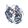
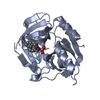
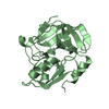
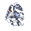
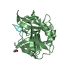
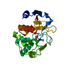
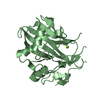
- PDB-6sf7: Atomic resolution structure of SplF protease from Staphylococcus ... -
+ Open data

- Basic information
| Entry | Database: PDB / ID: 6sf7 | ||||||
|---|---|---|---|---|---|---|---|
| Title | Atomic resolution structure of SplF protease from Staphylococcus aureus | ||||||
 Components Components | Serine protease SplF | ||||||
 Keywords Keywords |  HYDROLASE / Virulence factor / protease / bacterial HYDROLASE / Virulence factor / protease / bacterial | ||||||
| Function / homology |  Function and homology informationHydrolases; Acting on peptide bonds (peptidases); Serine endopeptidases / serine-type peptidase activity / extracellular region Function and homology informationHydrolases; Acting on peptide bonds (peptidases); Serine endopeptidases / serine-type peptidase activity / extracellular regionSimilarity search - Function | ||||||
| Biological species |  Staphylococcus aureus subsp. aureus USA300 (bacteria) Staphylococcus aureus subsp. aureus USA300 (bacteria) | ||||||
| Method | X-RAY DIFFRACTION / MOLECULAR REPLACEMENT / Resolution: 1.7 Å | ||||||
 Authors Authors | Golik, P. / Stach, N. / Karim, A. / Dubin, G. | ||||||
 Citation Citation | Journal: Int J Mol Sci / Year: 2021 Title: Structural Determinants of Substrate Specificity of SplF Protease from Staphylococcus aureus . Authors: Stach, N. / Karim, A. / Golik, P. / Kitel, R. / Pustelny, K. / Gruba, N. / Groborz, K. / Jankowska, U. / Kedracka-Krok, S. / Wladyka, B. / Drag, M. / Lesner, A. / Dubin, G. | ||||||
| History |
|
- Structure visualization
| Structure viewer | Molecule: MolmilJmol/JSmol |
|---|
- Downloads & links
-Download
| PDBx/mmCIF format | 6sf7.cif.gz | 315.6 KB | Display | PDBx/mmCIF format |
|---|---|---|---|---|
| PDB format | pdb6sf7.ent.gz | 256.7 KB | Display | PDB format |
| PDBx/mmJSON format | 6sf7.json.gz | Tree view | PDBx/mmJSON format | |
| Others |  Other downloads Other downloads |
-Validation report
| Arichive directory | https://data.pdbj.org/pub/pdb/validation_reports/sf/6sf7ftp://data.pdbj.org/pub/pdb/validation_reports/sf/6sf7 | HTTPS FTP |
|---|
-Related structure data
| Related structure data |  4vidS S: Starting model for refinement |
|---|---|
| Similar structure data |

-Links
 PDBj
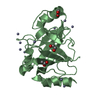
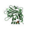
PDBj- Assembly
| Deposited unit | 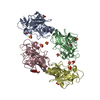
| ||||||||
|---|---|---|---|---|---|---|---|---|---|
| 1 | 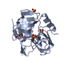
| ||||||||
| 2 | 
| ||||||||
| 3 | 
| ||||||||
| 4 | 
| ||||||||
| Unit cell |
|
-Components
-Protein , 1 types, 4 molecules ABCD
| #1: Protein | Mass: 22045.727 Da / Num. of mol.: 4 Source method: isolated from a genetically manipulated source Source: (gene. exp.) Staphylococcus aureus subsp. aureus USA300 (bacteria)Gene: splF, SAUSA300_1753 / Production host: Escherichia coli (E. coli)References: UniProt: Q2FFT4, Hydrolases; Acting on peptide bonds (peptidases); Serine endopeptidases |
|---|
-Non-polymers , 5 types, 469 molecules 


| #2: Chemical | ChemComp-PEG / Diethylene glycol Mass: 106.120 Da / Num. of mol.: 1 / Source method: obtained synthetically / Formula: C4H10O3 Mass: 106.120 Da / Num. of mol.: 1 / Source method: obtained synthetically / Formula: C4H10O3 | ||||||
|---|---|---|---|---|---|---|---|
| #3: Chemical | ChemComp-SO4 / Sulfate Mass: 96.063 Da / Num. of mol.: 14 / Source method: obtained synthetically / Formula: SO4 Mass: 96.063 Da / Num. of mol.: 14 / Source method: obtained synthetically / Formula: SO4#4: Chemical | Ethylene glycol Mass: 62.068 Da / Num. of mol.: 3 / Source method: obtained synthetically / Formula: C2H6O2 Mass: 62.068 Da / Num. of mol.: 3 / Source method: obtained synthetically / Formula: C2H6O2#5: Chemical | ChemComp-PG4 / | Polyethylene glycol Mass: 194.226 Da / Num. of mol.: 1 / Source method: obtained synthetically / Formula: C8H18O5 / Comment: precipitant*YM Mass: 194.226 Da / Num. of mol.: 1 / Source method: obtained synthetically / Formula: C8H18O5 / Comment: precipitant*YM#6: Water | ChemComp-HOH / | WaterMass: 18.015 Da / Num. of mol.: 450 / Source method: isolated from a natural source / Formula: H2O |
-Details
| Has ligand of interest | N |
|---|
-Experimental details
-Experiment
| Experiment | Method: X-RAY DIFFRACTION / Number of used crystals: 1 |
|---|
- Sample preparation
| Crystal | Density Matthews: 2.31 Å3/Da / Density % sol: 46.7 % |
|---|---|
| Crystal grow | Temperature: 293 K / Method: vapor diffusion, sitting drop / Details: 32.5 % PEG 400, 0.3 M Ammonium Sulphate pH = 3.5 |
-Data collection
| Diffraction | Mean temperature: 100 K / Serial crystal experiment: N |
|---|---|
| Diffraction source | Source: ROTATING ANODE / Type: RIGAKU MICROMAX-007 HF / Wavelength: 1.54 Å |
| Detector | Type: RIGAKU RAXIS IV++ / Detector: IMAGE PLATE / Date: May 16, 2011 |
| Radiation | Protocol: SINGLE WAVELENGTH / Monochromatic (M) / Laue (L): M / Scattering type: x-ray |
| Radiation wavelength | Wavelength: 1.54 Å / Relative weight: 1 |
| Reflection | Resolution: 1.7→30 Å / Num. obs: 77687 / % possible obs: 92.6 % / Redundancy: 3.6 % / Rmerge(I) obs: 0.047 / Net I/σ(I): 6.4 |
| Reflection shell | Resolution: 1.7→1.79 Å / Redundancy: 3.7 % / Rmerge(I) obs: 0.11 / Mean I/σ(I) obs: 13.9 / Num. unique obs: 10882 / % possible all: 88.8 |
- Processing
| Software |
| |||||||||||||||||||||||||||||||||||||||||||||||||||||||||||||||||||||||||||||||||||||||||||||||||||||||||||||||||||||||||||||
|---|---|---|---|---|---|---|---|---|---|---|---|---|---|---|---|---|---|---|---|---|---|---|---|---|---|---|---|---|---|---|---|---|---|---|---|---|---|---|---|---|---|---|---|---|---|---|---|---|---|---|---|---|---|---|---|---|---|---|---|---|---|---|---|---|---|---|---|---|---|---|---|---|---|---|---|---|---|---|---|---|---|---|---|---|---|---|---|---|---|---|---|---|---|---|---|---|---|---|---|---|---|---|---|---|---|---|---|---|---|---|---|---|---|---|---|---|---|---|---|---|---|---|---|---|---|---|
| Refinement | Method to determine structure: MOLECULAR REPLACEMENT Starting model: 4VID Resolution: 1.7→29.84 Å / Cor.coef. Fo:Fc: 0.967 / Cor.coef. Fo:Fc free: 0.951 / SU B: 3.79 / SU ML: 0.063 / Cross valid method: THROUGHOUT / σ(F): 0 / ESU R: 0.11 / ESU R Free: 0.11 Details: U VALUES : WITH TLS ADDED HYDROGENS HAVE BEEN ADDED IN THE RIDING POSITIONS
| |||||||||||||||||||||||||||||||||||||||||||||||||||||||||||||||||||||||||||||||||||||||||||||||||||||||||||||||||||||||||||||
| Solvent computation | Ion probe radii: 0.8 Å / Shrinkage radii: 0.8 Å / VDW probe radii: 1.2 Å | |||||||||||||||||||||||||||||||||||||||||||||||||||||||||||||||||||||||||||||||||||||||||||||||||||||||||||||||||||||||||||||
| Displacement parameters | Biso max: 85.7 Å2 / Biso mean: 27.698 Å2 / Biso min: 13.73 Å2
| |||||||||||||||||||||||||||||||||||||||||||||||||||||||||||||||||||||||||||||||||||||||||||||||||||||||||||||||||||||||||||||
| Refinement step | Cycle: final / Resolution: 1.7→29.84 Å
| |||||||||||||||||||||||||||||||||||||||||||||||||||||||||||||||||||||||||||||||||||||||||||||||||||||||||||||||||||||||||||||
| Refine LS restraints |
| |||||||||||||||||||||||||||||||||||||||||||||||||||||||||||||||||||||||||||||||||||||||||||||||||||||||||||||||||||||||||||||
| LS refinement shell | Resolution: 1.7→1.744 Å / Rfactor Rfree error: 0 / Total num. of bins used: 20
| |||||||||||||||||||||||||||||||||||||||||||||||||||||||||||||||||||||||||||||||||||||||||||||||||||||||||||||||||||||||||||||
| Refinement TLS params. | Method: refined / Refine-ID: X-RAY DIFFRACTION
| |||||||||||||||||||||||||||||||||||||||||||||||||||||||||||||||||||||||||||||||||||||||||||||||||||||||||||||||||||||||||||||
| Refinement TLS group |
|
