 Movie
Movie Controller


+ Open data

- Basic information
| Entry | Database: PDB / ID: 6sar | ||||||
|---|---|---|---|---|---|---|---|
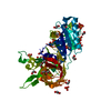
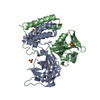
| Title | E coli BepA/YfgC | ||||||
 Components Components | Beta-barrel assembly-enhancing protease | ||||||
 Keywords Keywords |  CHAPERONE / outer membrane bam complex chaperone protease CHAPERONE / outer membrane bam complex chaperone protease | ||||||
| Function / homology |  Function and homology information Function and homology informationGram-negative-bacterium-type cell outer membrane assembly / Hydrolases; Acting on peptide bonds (peptidases) / protein disulfide isomerase activity / chaperone-mediated protein folding / proteolysis involved in protein catabolic process / metalloendopeptidase activity / outer membrane-bounded periplasmic space / zinc ion binding / membrane / metal ion bindingSimilarity search - Function | ||||||
| Biological species |  Escherichia coli (E. coli) Escherichia coli (E. coli) | ||||||
| Method | X-RAY DIFFRACTION / SYNCHROTRON / MAD / Resolution: 2.18 Å | ||||||
 Authors Authors | Lovering, A.L. / Cadby, I.T. | ||||||
 Citation Citation | Journal: J.Bacteriol. / Year: 2020 Title: Structure-Function Characterization of the Conserved Regulatory Mechanism of the Escherichia coli M48 Metalloprotease BepA. Authors: Bryant, J.A. / Cadby, I.T. / Chong, Z.S. / Boelter, G. / Sevastsyanovich, Y.R. / Morris, F.C. / Cunningham, A.F. / Kritikos, G. / Meek, R.W. / Banzhaf, M. / Chng, S.S. / Lovering, A.L. / Henderson, I.R. | ||||||
| History |
|
- Structure visualization
| Structure viewer | Molecule: MolmilJmol/JSmol |
|---|
- Downloads & links
-Download
| PDBx/mmCIF format | 6sar.cif.gz | 171.2 KB | Display | PDBx/mmCIF format |
|---|---|---|---|---|
| PDB format | pdb6sar.ent.gz | 135.7 KB | Display | PDB format |
| PDBx/mmJSON format | 6sar.json.gz | Tree view | PDBx/mmJSON format | |
| Others |  Other downloads Other downloads |
-Validation report
| Arichive directory | https://data.pdbj.org/pub/pdb/validation_reports/sa/6sarftp://data.pdbj.org/pub/pdb/validation_reports/sa/6sar | HTTPS FTP |
|---|
-Related structure data
| Similar structure data |
|---|


-Links
 PDBj
PDBj
- Assembly
| Deposited unit | 
| ||||||||
|---|---|---|---|---|---|---|---|---|---|
| 1 |
| ||||||||
| Unit cell |
|
-Components
| #1: Protein | Mass: 53962.766 Da / Num. of mol.: 1 Source method: isolated from a genetically manipulated source Source: (gene. exp.) Escherichia coli (strain K12) (bacteria)Strain: K12 / Gene: bepA, yfgC, b2494, JW2479 / Production host: Escherichia coli (E. coli)References: UniProt: P66948, Hydrolases; Acting on peptide bonds (peptidases) |
|---|---|
| #2: Chemical | ChemComp-ZN /   Mass: 65.409 Da / Num. of mol.: 1 / Source method: obtained synthetically / Formula: Zn Mass: 65.409 Da / Num. of mol.: 1 / Source method: obtained synthetically / Formula: Zn |
| #3: Chemical | ChemComp-SO4 / Sulfate  Mass: 96.063 Da / Num. of mol.: 1 / Source method: obtained synthetically / Formula: SO4 Mass: 96.063 Da / Num. of mol.: 1 / Source method: obtained synthetically / Formula: SO4 |
| #4: Water | ChemComp-HOH / Water Mass: 18.015 Da / Num. of mol.: 120 / Source method: isolated from a natural source / Formula: H2O Mass: 18.015 Da / Num. of mol.: 120 / Source method: isolated from a natural source / Formula: H2O |
| Has ligand of interest | N |
-Experimental details
-Experiment
| Experiment | Method: X-RAY DIFFRACTION / Number of used crystals: 1 |
|---|
- Sample preparation
| Crystal | Density Matthews: 2.36 Å3/Da / Density % sol: 47.92 % |
|---|---|
| Crystal grow | Temperature: 291 K / Method: vapor diffusion, sitting drop / pH: 7 / Details: 0.1 M Na HEPES, pH 7.0, and 8% w/v PEG 8,000 |
-Data collection
| Diffraction | Mean temperature: 100 K / Serial crystal experiment: N | ||||||||||||||||||||||||||||||
|---|---|---|---|---|---|---|---|---|---|---|---|---|---|---|---|---|---|---|---|---|---|---|---|---|---|---|---|---|---|---|---|
| Diffraction source | Source: SYNCHROTRON / Site: Diamond  / Beamline: I03 / Wavelength: 1.28274 Å / Beamline: I03 / Wavelength: 1.28274 Å | ||||||||||||||||||||||||||||||
| Detector | Type: DECTRIS PILATUS3 S 6M / Detector: PIXEL / Date: Dec 1, 2015 | ||||||||||||||||||||||||||||||
| Radiation | Protocol: SINGLE WAVELENGTH / Monochromatic (M) / Laue (L): M / Scattering type: x-ray | ||||||||||||||||||||||||||||||
| Radiation wavelength | Wavelength: 1.28274 Å / Relative weight: 1 | ||||||||||||||||||||||||||||||
| Reflection | Resolution: 2.18→77.02 Å / Num. obs: 27140 / % possible obs: 98.9 % / Redundancy: 16.5 % / CC1/2: 0.999 / Rmerge(I) obs: 0.116 / Rpim(I) all: 0.028 / Rrim(I) all: 0.119 / Net I/σ(I): 20.8 / Num. measured all: 448076 / Scaling rejects: 467 | ||||||||||||||||||||||||||||||
| Reflection shell | Diffraction-ID: 1
|
-Phasing
| Phasing | Method: MAD |
|---|
- Processing
| Software |
| ||||||||||||||||||||||||||||||||||||||||||||||||||||||||||||
|---|---|---|---|---|---|---|---|---|---|---|---|---|---|---|---|---|---|---|---|---|---|---|---|---|---|---|---|---|---|---|---|---|---|---|---|---|---|---|---|---|---|---|---|---|---|---|---|---|---|---|---|---|---|---|---|---|---|---|---|---|---|
| Refinement | Method to determine structure: MAD / Resolution: 2.18→77.02 Å / Cor.coef. Fo:Fc: 0.96 / Cor.coef. Fo:Fc free: 0.955 / SU B: 9.6 / SU ML: 0.118 / Cross valid method: THROUGHOUT / σ(F): 0 / ESU R: 0.199 / ESU R Free: 0.159 / Stereochemistry target values: MAXIMUM LIKELIHOOD Details: U VALUES : WITH TLS ADDED HYDROGENS HAVE BEEN ADDED IN THE RIDING POSITIONS
| ||||||||||||||||||||||||||||||||||||||||||||||||||||||||||||
| Solvent computation | Ion probe radii: 0.7 Å / Shrinkage radii: 0.7 Å / VDW probe radii: 1 Å / Solvent model: MASK | ||||||||||||||||||||||||||||||||||||||||||||||||||||||||||||
| Displacement parameters | Biso max: 116.01 Å2 / Biso mean: 47.398 Å2 / Biso min: 24.94 Å2
| ||||||||||||||||||||||||||||||||||||||||||||||||||||||||||||
| Refinement step | Cycle: final / Resolution: 2.18→77.02 Å
| ||||||||||||||||||||||||||||||||||||||||||||||||||||||||||||
| Refine LS restraints |
| ||||||||||||||||||||||||||||||||||||||||||||||||||||||||||||
| LS refinement shell | Resolution: 2.18→2.237 Å / Rfactor Rfree error: 0 / Total num. of bins used: 20
| ||||||||||||||||||||||||||||||||||||||||||||||||||||||||||||
| Refinement TLS params. | Method: refined / Origin x: 29.966 Å / Origin y: 48.42 Å / Origin z: 44.623 Å
|
